Biomedical Engineering Reference
In-Depth Information
Extensive physicochemical studies on RPR120535, a leading
product of the series, demonstrated the presence of characteristic
multi-lamellar bilayers, particles of 200 nm, with a surprising
periodicity of 80 A [28]. Unlike quaternary ammonium lipids, these
multi-lamellar particles are formed in the presence or absence of
any helper or additive lipid, such as DOPE. In an extensive study,
the structural polymorphism of DNA/RPR120535 complexes has
been studied by X-ray diffraction and cryo-electron microscopy
(see Fig. 1.6). Monovalent salts and temperature effects have been
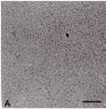
Figure 1.
RPR120535 and RPR120535/DNA complexes from zones A,
B, and C, visualized by light microscopy (TEM). (A) Cryo-TEM
micrographs of an aqueous solution of RPR120535 alone.
(B) Electron micrograph of uranyl acetate-stained complexes
from zone A (Charge ratio +/-: 0.3). Inset shows the same
complexes at higher magnification. (C) Complexes from zone
B (Charge ratio +/-: 1.65) observed by light microscopy.
(D) Same complexes as (C) stained with uranyl acetate. (E)
Cryo-TEM micrograph of RPR120535/DNA complexes from
zone C (Charge ratio +/-: 6). Inset shows the visualization
by cryo-TEM of the ordered domains in these complexes. (F)
Electron micrograph of uranyl acetate stained complexes from
zone C. (G) Cryo-phosphotungstate-TEM micrograph of the
same complexes. In this micrograph, the complexes seem to
have aggregated, because the thickest part of the vitrified film
allows them to be superimposed. The scale bar represents
100 nm in A, B inset, and D-G; 500 nm in B; and 10 micron in C.
Reproduced with permission [28].


Search WWH ::

Custom Search