Biomedical Engineering Reference
In-Depth Information
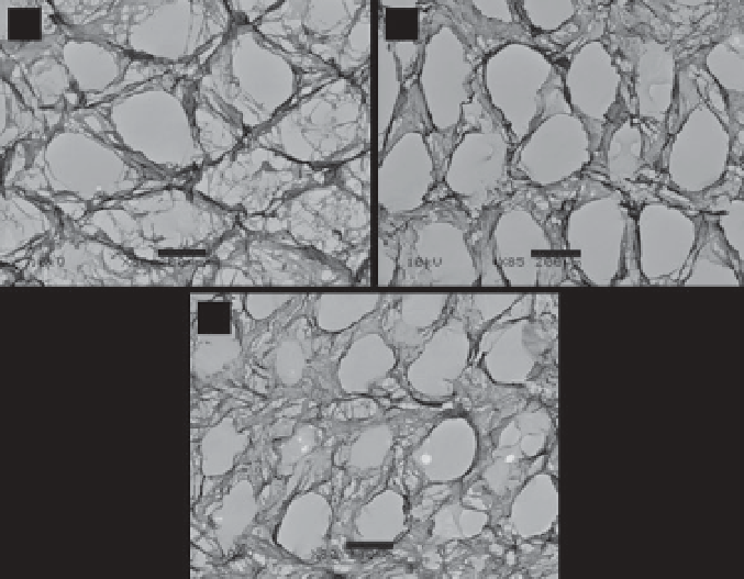
microscopy (SEM) demonstrates the porous structure of microscale and
nanoscale ECM fi bers (Figure 10.2). There is no obvious difference among
ECM-M, ECM-C and ECM-F scaffolds in terms of morphology. The geo-
metrical properties, porosity, interconnectivity, and nanoscaled fi brous
structure are meant to support cell proliferation and differentiation and
benefi t tissue regeneration [31, 32]. The cell nuclei, cell membrane and
F-actin are removed by a decellularization treatment. Removal of PLGA
mesh template is confi rmed by AIR-FTIR spectra which show the ester
carbonyl stretch at 1740 cm
-1
in the cell-ECM-PLGA complexes disappears
in the ECM scaffolds. Examination of the composition of ECM scaffolds
shows there is some difference among the compositional biomolecules
in ECM-M, ECM-C and ECM-F (Table 10.1). ECM-M consists of type I
collagen, type III collagen, fi bronectin, vitronectin, laminin, aggrecan,
decorin and biglycan. ECM-C consists of type I collagen, type III colla-
gen, fi bronectin, vitronectin, laminin, aggrecan, versican, decorin and
biglycan. ECM-F consists of type I collagen, type III collagen, fi bronectin,
vitronectin, laminin, decorin and biglycan. Although primary chondro-
cytes express type II collagen, the ECM-C does not contain type II collagen
because dedifferentiated passage 5 chondrocytes that do not express type
II collagen are used to prepare the ECM scaffolds. Therefore, the compo-
sition of the ECM scaffolds depends on the cell type and cell phenotype
used to prepare the scaffolds.
(
a
)
(
b
)
(
c
)
Figure 10.2
SEM image of ECM scaffolds prepared from MSCs (a), chondrocytes
(b) and fi broblasts (c). Scale bar = 200 μm.
Search WWH ::

Custom Search