Biomedical Engineering Reference
In-Depth Information
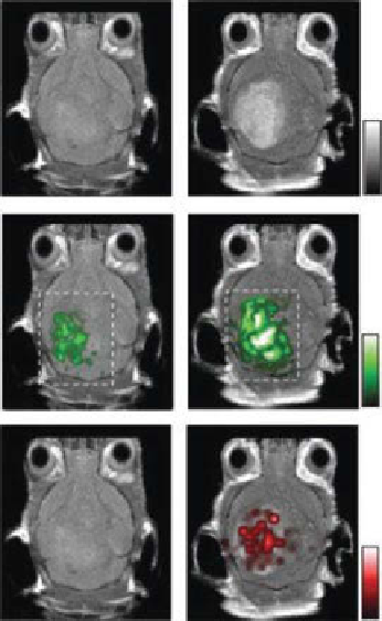
(a)
Before injection
After injection
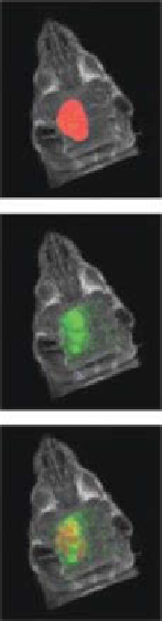
(b)
3D rendering
Max
0
Max
0
Max
0
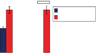
(c)
***
***
**
18
1.2
16
1.0
Before injection
After injection
14
12
0.8
10
0.6
8
6
0.4
4
0.2
2
0
0
MRI
Photoacoustic
Raman
figure 11.9
Triple-modality detection of brain tumors in living mice with magnetic
resonance imaging-photoacoustic imaging-Raman imaging (MPR) nanoparticle. (a) Two-
dimensional axial magnetic resonance imaging (MRi), photoacoustic, and Raman images. The
postinjection images of all three modalities showed clear tumor visualization (dashed boxed
outline highlighted the imaged area). (b) a three-dimensional (3D) rendering of magnetic reso-
nance images with the tumor segmented (light grey; top), an overlay of the 3D photoacoustic
images (light grey) over the MRi (middle), and overlay of MRi, the segmented tumor, and the
photoacoustic images (bottom) showing good colocalization of the photoacoustic signal with the
tumor. (c) Quantification of the signals in the tumor showing a significant increase in the MRi,
photoacoustic, and Raman signals after as compared to before the injection.
n
= 4 mice. Data
represent mean ± s.e.m. ***P<0.001, **P<0.01 (one-sided Student's
t
test.) au, arbitrary units.
(Reprinted with permission from Ref. [20]. © Macmillan Publishers Ltd.)


Search WWH ::

Custom Search