Biomedical Engineering Reference
In-Depth Information
(a)
(d)
PA+US
Agent #1
Agent #2
Water
Water
Agent #3
Rat skin
CT
1 cm
SLN
(b)
1 cm
10 mm
(e)
CT/PAT
CT/SPECT
CT/SPECT
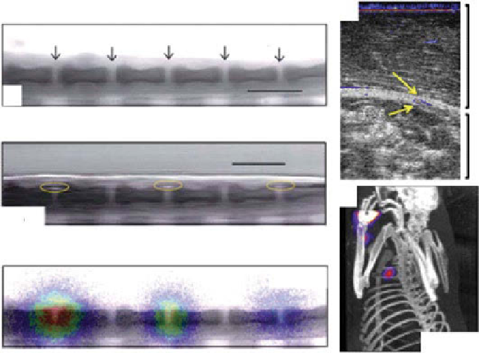
figure 10.15
Multimodal imaging of radiolabeled optical absorbents in small animals. (a)
MicroCT image of a sagittal section of joints in a rat tail after intra-articular injection of gold
nanorods labeled with
125
I. (b) overlaid PA and CT image of the joint section. (c) overlaid CT
and SPECT image of the joint. (d) overlaid PA and US image of a sentinel lymph node after
intradermal injection of methylene blue. (e) SPECT/CT projection image of the rat axillary
region acquired at 1 h postinjection of
125
I methylene blue. (reprinted with permission from
refs. [150, 151]. © SPIE and © Elsevier.)
(i.e., bone) were clearly shown in the microCT image (Fig. 10.15b). In Figure 10.15c,
SPECT image was overlaid with the microCT image visualizing both the distribution
of contrast agent and the hard tissue morphologies. As one can see, the contrast of
nuclear imaging was stronger, whereas the resolution of PAT was better to show
microstructures. As the second approach, MB was radiolabeled with
125
I as a PA and
nuclear lymph node tracer [150]. PA and nuclear imaging were performed to map
axillary lymph nodes in small animals
in vivo
. An overlaid PA and US image
(Fig. 10.15d) provides both functional (MB uptake in the sentinel lymph node) and
structural information of surrounding tissues, respectively. The SPECT image clearly
visualizes the localized sentinel lymph node uptaken by
125
I MB in the axillary region
at 1 h postinjection, while the CT image shows the surrounding structures (Fig. 10.15e).
10.5.8
organic dyes
Highly optically absorbing organic dyes (i.e., having high nonradiative quantum
yield) have been widely applied as a PA contrast agent to various preclinical and
clinical applications. ICg [85], MB [83], and Evans blue (EB) [152, 153] have
Search WWH ::

Custom Search