Biomedical Engineering Reference
In-Depth Information
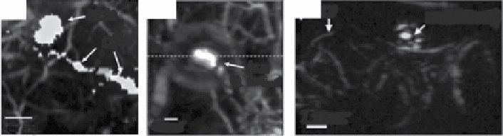
(a)
(b)
(c)
LV
SLN
SLN with EB
LV
a
SLN
with MB
1 mm
2 mm
5 mm
figure 10.16
In vivo
PA images of sentinel lymph nodes (SLns) in small animals after
intradermal injection of (a) indocyanine green (ICg), (b) methylene blue (MB), and (c) Evans
blue (EB). (reprinted with permission from refs. [83, 85, 153]. © SPIE, © The American
Association of Physicists Medicine, © radiological Society of north America.)
already been used in clinics, which is the strong advantage of using these organic
dyes. The molar extinction coefficients of ICg, MB, and EB are 1.5 × 10
5
, 7.4 × 10
4
,
and 7.8 × 10
4
cm
−1
M
−1
, respectively. The peak absorption wavelengths of ICg and
MB are 790 and 667 nm, respectively, within the nIr window (650-900 nm),
while that of EB is 626 nm. However, the drawbacks of these dyes include the
short circulation time in a bloodstream, smaller optical absorption cross sections,
and difficult surface modification. ICg, MB, and EB have been successfully used for
noninvasive mapping of sentinel lymph nodes in small animals
in vivo
(Fig. 10.16).
10.5.9
fluorescence Proteins [154]
Imaging FL proteins using FL microscopy plays an important role in biological
researches. As aforementioned, however, the applications of FL proteins are limited
because of the shallow imaging depth of optical imaging modalities. Some FL pro-
teins having small fluorescent quantum yields (i.e., high nonradiative quantum yield)
can be great contenders as PA contrast agents. Since PAT can achieve deep tissue
imaging, molecular PAT combined with FL proteins has great potential in biological
studies. For the first time, the expression of mCherry in an adult transgenic zebra fish
was photoacoustically mapped
in vivo
[155]. However, visible optical wavelengths,
where the peak optical absorption of mCherry is, were used, and the imaging
depth was consequently limited in this approach. recently, Filonov
et al
. have
developed an nIr FL protein, bacteriophytochrome-based nIr FL proteins (named
irFP), as a PA probe [154]. irFP possesses a very high molar extinction coefficient
of 1,05,000 cm
−1
M
−1
and a low fluorescent quantum yield of 6%. To prove the strong
PA contrast of irFP, the PA signals measured from purified irFP, E2-Crimson,
mneptune, mKate2, eqFP670, and TagrFP657 proteins were compared with lysed
oxygenated blood
in vitro
(Fig. 10.17a). The PA amplitude of irFP at 680 nm was
higher than that at 600 nm and was the highest among all proteins. Figure 10.17
shows the spectrally decoded PA images of a mouse mammary gland tumor
in vivo
without (Fig. 10.17b) and with (Fig. 10.17c) the tissue overlay. The boundary of the
tumor positioned at a depth of 4 mm in tissue was clearly identified.
Search WWH ::

Custom Search