Biomedical Engineering Reference
In-Depth Information
5.2.3
fluorescent proteins and enzymatic approach
fluorescent proteins are widely used in cell biology and in the study of transgenic
animals, because they can be genetically targeted to a specific molecule of interest
[61]. One major advantage of using fluorescent proteins as the contrast agent for
OA imaging is that they overcome the limitations of conventional fluorescence
microscopy and allow imaging of gene expression in much deeper tissues [62].
It has been reported that msOT can achieve tissue penetration of several milli-
meters (potentially centimeters) with a resolution of 20-100 µm, which remains
constant as a function of depth and depends only on the ultrasonic detector
characteristics [20]. This technique was capable of visualizing fluorescent pro-
teins in small living organisms such as the zebra fish,
Drosophila melanogaster
pupae, and adult zebra fish for high-resolution morphological and functional
observations (fig. 5.5).
(a)
1
(b)
(c)
DM
B
N
O
S
P
0.5 mm
H
HM
2 mm
0
(d)
(e)
600 μm
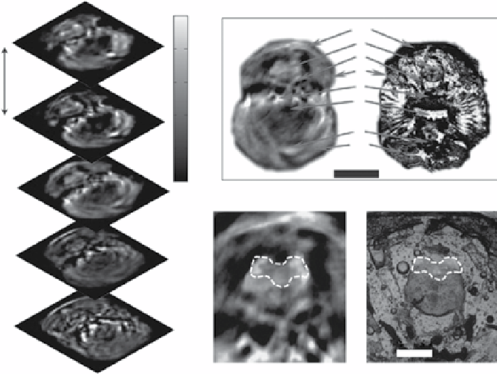
figure 5.5
Three-dimensional
in vivo
imaging through the brain of an adult mcherry-
expressing transgenic zebrafish. (a) five transverse optoacoustic image slices through the
hindbrain area at the level of crista cerebellaris of a living zebrafish taken at 585 nm. (b,c)
example of an imaged slice (b) and its corresponding histological section (c). Dm, dorsal fin
musculature; B, hindbrain; N, lateral line nerve; O, operculum; s, skull bones; P, pharynx; H,
heart; Hm, hypobranchial musculature. (d) msOT image of the brain with mcherry expres-
sion shown in the area outlined with a dashed line; and (e) corresponding epi-fluorescent
histology of the fish showing the same area of mcherry expression. (Adapted with permission
from Ref.[20]. Nature Publishing group).
Search WWH ::

Custom Search