Biomedical Engineering Reference
In-Depth Information
(a)
(b)
(c)
(d)
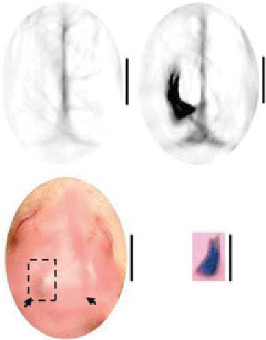
figure 5.6
Optoacoustic images at 650 nm before and after the injection of X-gal (planes
a
and
b
, respectively). Plane
c
shows the photograph of the rat head following X-gal injection
and just prior to imaging. The two arrows indicate the position of injection of X-gal. Plane
d
shows the underside of the dissected scalp in the rectangular region indicated in plane
c
, show-
ing the X-gal stained tumor, which is dark blue in color (
d
). The line scale represents 5 mm.
(Reproduced with permission from Ref. [63]. © sPIe-International society for Optical
engineers.)
exogenous nonplasmonic contrast agents can be used for enhancement of OA
imaging with gene expression. The study [63] first demonstrated the capability of
OAT for reporter gene imaging. Rats inoculated with 9L/lacZ gliosarcoma tumor
cells were imaged with OAT before and after injection of X-gal, an optically trans-
parent lactose-like substrate, which yields a stable dark-blue product following the
cleavage of the glycosidic linkage by β-galactosidase (the protein encoded by lacZ).
figure 5.6 shows the OA image prior to X-gal injection (fig. 5.6), and the tumor is
clearly seen after X-gal administration (fig. 5.6). A spatial resolution of around
400 µm, as well as a sensitivity of 0.5 μm, was achieved. No signal upregulation was
observed at the right-side injection point, which served as a negative control.
5.3 exogeNous NoNplasmoNic coNtrast ageNts
for optoacoustic imagiNg
Although the progress made to date by OAT and imaging with endogenous markers
such as hemoglobin and melanin has been significant, the use of exogenous contrast
agents has further increased the benefits of this imaging modality by enhancing the
Search WWH ::

Custom Search