Biomedical Engineering Reference
In-Depth Information
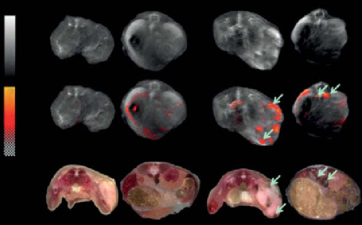
optoacoustic tomographic (msOT) imaging of melanin-containing tumors, the
authors of Reference [54] were able to generate an image-processing algorithm that
allowed to visualize the presence and distribution of melanin not only on the surface
but also deep within the mouse body. With this technique, near-surface A549 and
Pc-3 tumors, as well as Pc-3-RfP lymph node metastases (fig. 5.4), which, in a
recent study, have been shown to be preferentially colonized versus the primary
tumor [57], could be detected in live mice injected with melanin-rVAcV.
compared with fluorescent proteins (see following text), melanin can certainly
not be used for subcellular protein distribution and interaction analysis, but it has the
advantage of being visible via noninvasive deep tissue imaging with OAT imaging.
The detection of fluorescent proteins is probably more sensitive than melanin in
terms of generated signal per molecule, but melanin is produced enzymatically,
which allows relatively fast detection (about 18 h post viral cell infection) with
increasing intensity over time [54]. Another advantage of melanin versus fluorescent
proteins is the extreme stability of melanin [58]. These data have shown that gene-
evoked melanin production has a strong potential to become widely used in surgery
and endoscopy with OA imaging. finally, the ubiquitous presence of melanin in all
kingdoms of life suggests that introduction of melanin synthesis as a diagnostic/
theranostic marker will be possible in most species [59] and should also translate into
clinical settings [54]. However, the process of melanin production in transfected cells
is thought to be toxic [60]. future studies must investigate different ways of melanin
delivery and focus on
in vivo
applications of this technology.
Control-rVACV
Melanin-rVACV
4.5
-1
5
0
LN2
LN2
LN1
LN1
Tumor
Tumor
figure 5.4
Optoacoustic imaging of melanin-rVAcV in live mice. multispectral optoacous-
tic tomography (msOT) of Pc-3-RfP tumor and lymph node metastasis (LN1 and LN2) bearing
live mice 14 days after rVAcV injection. (Top) Background optoacoustic images taken at 850 nm
excitation wavelength. (middle) melanin spatial distribution (pseudocolor) is overlaid on the
background images using a variable transparencies function (Bottom) As indicated by the
arrows
.
(With permission from Ref. [54]. © PNAs.) (
See insert for color representation of the figure.)
Search WWH ::

Custom Search