Biomedical Engineering Reference
In-Depth Information
after the implantation (
p
< 0.05). For the applications of calcium phosphate coating to den-
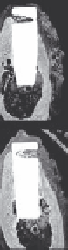
tal implants, implantation tests of coated Ti have been reported. Figure 7.33 shows optical
micrographs of the interface between the ACP-coated and noncoated (control) CP Ti cylin-
ders and bones 2, 4, 8, and 12 weeks after implantation in mandibles of beagle dogs.
(86)
BIC,
which is the ratio of the surface area of the implant in direct contact with bone to total sur-
face area, is shown in Figure 7.34. The percentage of BIC for the ACP-coated CP Ti cylinders
was greater than that for the noncoated cylinders 8 to 12 weeks after implantation.
(86)
In the evaluation of calcium phosphate coatings, both the amorphous and crystalline
phases have been employed. Crystalline calcium phosphate coating with a thickness of
1 μm on Ti substrates implanted into the backs of New Zealand white rabbits was still
detected 12 weeks after implantation, while ACP coating with a thickness of 4 μm dis-
appeared and then a crystalline carbonate apatite layer was precipitated.
(83)
A crystalline
calcium phosphate coating with a thickness of 0.1 μm remained 3 weeks after implantation
in the femur of goats, owing to the high stability of crystalline calcium phosphate thin film
under biological conditions.
(84)
Cross sections of Ti implants with amorphous and crystalline calcium phosphate coat-
ings after implantation in the femurs of Japanese white rabbits for 4 weeks are shown in
Figure 7.35a and b, respectively. The ACP coating was prepared by RF magnetron sput-
tering and the crystalline phase was obtained by the postheat treatment of as-sputtered
OAp in air for 7.2 ks. Direct contact between implants and new bones was microscopi-
cally observed for both types of implants. The crystalline calcium phosphate coating still
existed at the interface between Ti and new bone 4 weeks after implantation, while no
layer was detected on the ACP-coated Ti implant. The high bioresorbability of ACP coat-
ing can be confirmed in bones similar to that in SBF. The elution of calcium and phosphate
ions from bioceramic coatings might have affected the bone forming ability. Vapor deposi-
tion methods are suitable to control the composition and phase of the bioceramic coatings.
The effect of eluted ions, not only calcium and phosphate ions but also other ions such as
silicon or zinc, should be studied using calcium phosphate coatings prepared by vapor
deposition.
The bone forming ability of implants may be closely related to the initial interaction
with biomolecules. The immobilization of biomolecules such as bisphosphonates
(47,85,87)
and fibronectin(88)
(88)
on the surface of calcium phosphate-coated Ti by PVD has been studied.
2 weeks
4 weeks
8 weeks
12 weeks
ACP
coated
Control
FIGURE 7.33
Optical micrographs of interface between ACP-coated and noncoated (control) CP Ti cylinders and bones 2, 4, 8,
and 12 weeks after implantation in mandibles in beagle dogs.

Search WWH ::

Custom Search