Chemistry Reference
In-Depth Information
Gd
3+
Functional molecule
e.g. FA, AA, or OA
1. CHCl
3
, untrasonic
2. Gd
3+
18
F
-
3. New surface ligand
4.
18
F
-
UCNPs
UCNPs
OM:
H
2
N
: OM
R-COOH:
HOOC
N
N
N
N
: R-COOH
NH
2
FA
N
HOOC
NH
O
:
18
F
-
O
OA
HOOC
: Gd
3+
HOOC
NH
2
: Er
3+
AA
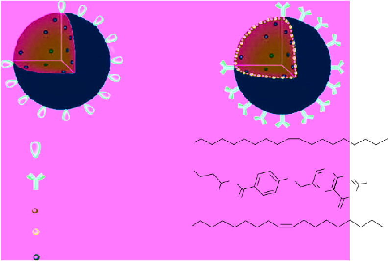
ScheMe 16.10
Modification of UCNPs with Gd
3+
and F
-
ions and carboxylic acid-bearing surface ligands. Reprinted with permission
from Ref. [80]. Copyright 2011 American Chemical Society.
Surface ligand exchange can be used to solubilise NaYF
4
particles (UCNPs) as well as introduce other ions and targeting
ligands. In this report, Gd
3+
ions,
18
F
-
ions, and aminocaproic acid (AA) are introduced during sonication [80]. The ions
exchange on the surface and are subsequently stabilised by the acid ligand (Scheme 16.10). The
r
1
per Gd(III) ion was found
to be 28 mM
-1
s
-1
at 3 T, 38 °C.
In vivo
imaging in mice was undertaken using AA-Gd-UCL-NPs [80]. Rapid accumulation in the liver and spleen was
observed as expected in the UCL image (Figure 16.21).
T
1
-weighted MR imaging showed 30% enhancements in the liver
and spleen 10 min. after injection.
18
F-AA-Gd-UCL-NPs were used for
in vivo
PET imaging and showed rapid uptake by the
liver and spleen and very low uptake by other organs, including bone.
16.5.3
theranostic applications of ucl-nps
Two nanocomposites are described where UCL nanoparticles are combined with iron oxide particles in a polymeric shell and
either a squaraine dye (SQ, Figure 16.22, synthesised as in Ref. [81]) or DOX was added [82]. The polymers used were
poly(styrene block allyl alcohol) and polyvinyl alcohol. Iron oxide particles of 6-8 nm in size were incorporated into the
nanocomposite using a microemulsion method. The overall
r
2
of the particles was found to be 84 mM
-1
s
-1
, with the diameter
of the nanoparticles being about ~200 nm. pH-dependent release of DOX was observed, with more DOX being released at
lower pH. Cell toxicity studies showed that UCL-DOX was slightly less toxic than free DOX. Magnetic targeting was
employed to localise the particles in cell culture. Uptake of UCL-DOX was higher in cells growing near the magnet and cell
viability was lower.
4 T1 cells were labelled with the UCL-SQ nanoparticles for 24 h and injected into mice. After 10 days, the UCL could
still be observed as well as the down conversion fluorescence from the SQ. Between 0 and 5 days, the MR signal decreased,
possibly due to tumour growth.
Biodistribution of the UCL-SQ particles showed that the particles localised quickly in the liver (within 2 h).
T
2
weighted
MR imaging showed darkening of the liver, spleen, and lung, but no change in the other major organs.
To take advantage of the photothermal therapeutic properties of Au nanoparticles, UCL nanoparticles were combined
with iron oxide particles and coated with a thin shell of gold via a seed-mediated procedure [83]. The final particles consisted
of cube-like structures with a diameter of approximately 200 nm, with ratio of Y/Fe/Au of 100:37:4. When irradiated with
808 nm laser light at 1 W/cm
2
, the temperature of the solution increased rapidly, indicating that the UCL emission is unper-
turbed in this nanocomposite.