Chemistry Reference
In-Depth Information
(a)
CO
2
H
I
I
HO
2
C
NH
2
I
O
(b)
O
H
O
N
N
O
O
n
O
O
fIGure 16.19
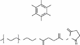
Structures of (a) 5-amino-2,4,6-triiodoisophthalic acid (AIPA) and (b) SEMG.
OH
O
HO
O
O
OH
HO
OH
O
HO
O
OH
HO
OH
O
O
OH
HO
OH
O
OH
O
OH
OH
OH
OH
O
O
O
HO
fIGure 16.20
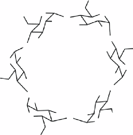
Structure of α-cyclodextrin, commonly used to host hydrophobic guests.
Solutions of the particles were injected into the subcutaneous tissue and intramuscular tissue of rats and visualised using
both UCL and CT. The UCL signal was weak but observable for the particles injected subcutaneously. Further, using control
solutions
in vitro
, it was found that the rare earth elements of the UCL particles contributed to the CT contrast.
16.5.2
biodistribution (ucl-pet)
UCL nanoparticles (UCL-NPs) can be synthesised from rare earth salts by solvothermal, thermal decomposition, and
hydrothermal methods [79]. The resulting hydrophobic UCL-NPs can be solubilised in aqueous solutions using
alpha
-
cyclodextrin (α-CD, Figure 16.20) to make CD-UCL-NPs [79]. Further, the probes can be used for PET imaging by incor-
porating
18
F for biodistribution and
in vivo
imaging.
The size of the particles prepared by the solvothermal and thermal decomposition methods was approximately ~18 nm.
Addition of α-CD combined with shaking solubilised the particles. Larger particles prepared using the hydrothermal method
(400 nm) were also solubilised using this method.
In vivo
UCL imaging of tail vein-injected CD-UCL-NPs showed the nanoparticles are taken up largely by the liver and
spleen, as would be expected for nanoparticles of this size. For lymphatic imaging, UCL-NPs were injected into the mouse
claw with expected lymphatic drainage to the oxter nodes as observed by UCL imaging.
18
F-labelled particles were generated by ligand exchange on the particle surface and used for
in vivo
microPET imaging
and
ex vivo
biodistribution. The particles were found to quickly accumulate in the liver and spleen. Uptake by other organs
was minimal. Because bone can take up free F
-
ions, the amount of
18
F in the bone was investigated and found to be very
low, supporting claims of the biostability of these particles.


