Chemistry Reference
In-Depth Information
OH
O
O
HO
O
NH
2
n
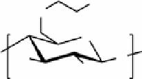
fIGure 16.15
Glycol chitosan (GC) is a polysaccharide composed of β-(1-4)-linked D-glucosamine subunits where one alcohol has
been modified with a glycol (-CH
2
CH
2
OH) group.
These GC particles were loaded with DOX. The drug release was evaluated at pH 7.4 and 6.4. At pH 6.4, drug release reached
a plateau within 6 h, whereas at pH 7.4, complete drug release was not reached for 48 h.
To track the particles
in vivo
,
131
I-labelled nanoparticles were used. Scintigraphic images of tumour-bearing mice show
GC3 particles, or those prepared with the highest wt% NAcHis targeted the tumour well while the other formulations (GC1
and GC2) did not. This result is in contrast to the NIRF images, which were comparable for all three compositions, but high-
est intensity in the tumour region was found for GC3.
Ex vivo
imaging showed high fluorescence in the tumour tissue and
some fluorescence in the liver, spleen, and kidneys.
The
in vivo
images for the DOX-loaded GCs varied dramatically. Although the scintigraphic images were very similar for
GC1 and GC2, the GC1 DOX formulation localised to a much larger extent in the tumour according NIRFI.
Ex vivo
NIRF
imaging shows fluorescence in the lung, liver, kidney, with the highest intensity in the tumour.
In order to test the drug delivery capability, HT29 tumour-bearing mice were treated with DOX, GC1, and DOX-GC1
every 3 days (5 mg dox/kg). Tumour volume was monitored over 16 days and growth was inhibited best by DOX-GC1.
To generate a PET-NIRFI-MRI trimodal agent, liposomes were formed with gadolinium labels (GdDOTA-DSPE) for
MRI, IRDye for NIRFI, and empty chelates (DOTA-DSPE) for PET nuclei (Figure 16.16). The liposomes were formed by
dissolving all the components in chloroform, followed by lipid film hydration and extrusion [66]. The authors demonstrated
that the DOTA-DSPE chelates could bind
64
Cu ions for PET imaging.
99m
Tc BMEDA was formed by reduction of NaTcO
4
with SnCl
2
in the presence of BMEDA and loaded into the preformed Gd-liposomes.
In vivo
NIRFI showed superior accumulation in the tumour of the liposomes when compared to the free NIR dye. The
excised tumour showed significantly more uptake than the other organs. Planar gamma camera images were obtained for
mice injected with
99m
Tc-labelled liposomes and showed accumulation in the tumour and fast clearance of the remainder. 3D
tomography and projection images from the microPET imaging of the
64
Cu labelled liposomes shows local retention and
micro-distribution of the radioactivity within the tumour.
16.4.3
tumour targeting using nIr-pet bimodal nanoparticle Systems
Glycol chitosan was modified with 5-β-cholanic acid (Figure 16.17) and Cy5.5 and the bladder cancer targeting peptide
CSNRDARRC [68]. This polymer was sonicated in the presence of iron oxide nanocubes, which became non-covalently
incorporated within the polymer matrix. The average diameter of the final particles was found to be 480 nm.
In vitro
cellular targeting was investigated using K9TCC bladder cancer cells incubated with the nanocubes for 3 h.
Confocal microscopy showed nanoparticles bound to the K9TCC cells. Lower concentration of nanocubes corresponds to
lower NIRF and decreased negative MR contrast, as expected.
NIRF imaging was utilised to evaluate the contrast effect of the modified nanocubes
in vivo
. Images of tumour-bearing
mice clearly indicated strong NIRF intensity at the tumour 24 h post administration of the nanoparticles.
Ex vivo
images
showed much brighter NIRF in the tumour than liver, spleen, and kidneys. The lung and heart showed negligible NIRF. This
result supports active targeting and retention of the nanocubes at tumours in mice. The results might be explained by both
passive accumulation of nanoparticles through EPR effects and active targeting of nanoparticles by specific binding effect
of CSNRDARRC peptide to bladder cancer. No MR imaging of the mice was reported.
Ferritin is a protein composed of 24 subunits that self-assemble into a nanostructure12 nm in size with an internal cavity
of 8 nm [69]. The subunit proteins may be chemically or genetically modified and may be assembled to encapsulate metal
ions within the hollow cavity [70-72]. In this report, ferritin was genetically modified with RGD, the integrin-targeting pep-
tide, and chemically modified with Cy5.5 [73]. The RGD-ferritin and Cy5.5-ferritin assemblies were combined, dissociated
at pH 2, and then reconstituted at pH 7.4 in the presence of
64
Cu
2+
. This procedure scrambles the ferritin components, and
re-assembly results in ferritin species with both RGD and Cy5.5 on the outer surface and a payload of
64
Cu
2+
ions encapsu-
lated within the cavity.
PET and NIRF images show the labelled ferritin is taken up by the tumour
in vivo
. A decrease in levels is observed bet-
ween 24 and 40 h in the PET images, while NIRF images show a decrease between 4 and 24 h. Administration of a blocking