Chemistry Reference
In-Depth Information
N
R
N+
R
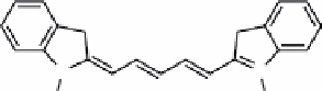
fIGure 16.4
The conjugated structure of cyanine dyes imparts their photophysical attributes. The R groups are alkyl chains with
or without functional groups such as an NHS ester or maleimide. The '5' in Cy5 refers to the number of carbon atoms bridging the two
heterocyclic indole groups.
1. FeCl
3
(H
2
O)
6
, FeCl
3
(H
2
O)
4
, 30% NH
4
OH, heat
2. 5M NaOH, H
2
O, 24 h, RT
OH
O
O
O
HO
HO
O
Cl
O
O
OH
O
O
NH
2
3. NH
4
OH, 24 h, 37°C
O
HO
HO
O
HO
OH
O
O
Crosslinked
dextran
O
n
Dextran
O
HO
Cl
ScheMe 16.5
Dextran coating and cross-linking on SPION surface results in -OH, -NH
2
, and -Cl reactive groups. Adapted from
refs. [30, 31].
16.3.1
cell tracking using nIr-MrI bimodal agents
The monitoring of B-cell depletion is a critical need in immunotherapy because it can be used to evaluate the biological
response to and efficacy of immunotherapeutics. In this report, dextran-coated SPIONs (Scheme 16.5) were labelled with a
NIR dye AlexaFluor680 (Invitrogen) [28]. B-cells were harvested from mice and labelled with the nanoparticles and a lipo-
philic dye, CellVue NIR 815 (Polysciences, Inc.).
NIR815-labelled B-cells with and without SPIONs were administered to mice and tracked using MR and NIR fluores-
cence imaging. The mice were treated with either PBS or antiCD79. NIRF imaging showed depletion of B cells treated with
NIR815, but not the cells labelled only with the SPIONs. Although the expression levels of several cell surface markers,
including CD40, CD80, CD86m, and MHC11, did not change when the cells were labelled with the nanoparticles, it appears
that labelling with the SPIONs had an effect on the ability of antiCD79 to interact with the B-cells.
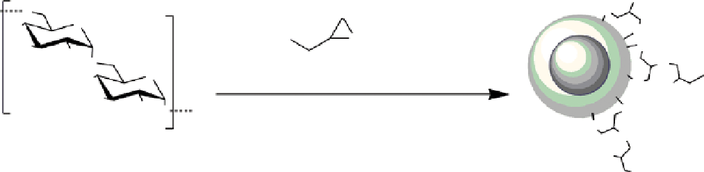
In some systems, NIR fluorescence is an intrinsic property. Kim et al. report the labelling of therapeutic dendritic cells
with a nanogel comprised of cross-linked manganese ferrite nanoparticles [32]. The MnFe
2
O
4
particles are coated with poly-
glutamic acid (PGA), then treated with polylysine (PLL), which electrostatically adsorbs to the PGA layer (Figure 16.5).
Cross-linking to form the nanogel is performed using glutaraldehyde (Figure 16.5) that forms a series of imine bonds bet-
ween the side chain amines of lysine. A consequence of this cross-linking is the formation of a Schiff base that produces a
surprisingly strong autofluorescence due to the conjugated bonds.
Dendritic cells were labelled with this nanogel and injected into nude mice. The cells were found to migrate to the lymph
nodes. A darkening in the MR image of the mouse was observed, indicating that the nanogel is an effective T
2
MRI contrast
agent
in vivo
. The lymph nodes were detected using NIRFI, further indicating that the nanogel is effective for
in vivo
imaging. The excised organs were imaged and exhibited strong NIR fluorescence.
Quantum dots (QDs) are nanoparticles based on semiconducting materials. Due to their nanometer size, their electronic
properties fall between those of bulk materials and those of discrete molecules. Semiconductor materials that are free of
heavy metals, such as nanoscale silicon, are being investigated as replacement materials for the development of quantum
dots due to the toxicity of cadmium, lead, and gallium commonly found in QDs [33, 34].
A magnetofluorescent probe with luminescence and superparamagnetic properties has been produced by combining
silicon-based quantum dots (SiQDs) and SPIONs in micelles [35]. SiQDs were synthesised by CO
2
laser pyrolysis of
silane (SiH
4
) in an aerosol reactor and found to be ~4 nm in diameter [36]. These particles were co-encapsulated in a
lipid micelle of phospholipids (Figure 16.6, 1,2-distearoyl-sn-glycero-3-phosphoethanolamine-N-[amino(polyethylene
glycol)-2000]; DSPE-PEG 2000; n ~ 142) with iron oxide particles (5-9 nm) to give a ratio by mass of Si:Fe
3
O
4
= 80:1.
The micelle size varied from 50-100 nm. If the amount of Fe
3
O
4
was increased, quenching of the SiQD fluorescence was
observed.