Chemistry Reference
In-Depth Information
O
OH
O
OH
OH
H
OO
OH
O
O
OH
NH
2
fIGure 16.3
Structure of doxorubicin (DOX), a widely used cancer therapeutic. In biological conditions, the -NH
2
group is protonated
to -NH
3
+
, giving an overall charge of +1 to the molecule.
O
O
HO
HO
O
O
HO
HO
N
N
N
N
H
2
NR
N
N
OH
OH
O
O
S
N
HN
C
C
S
HN
R
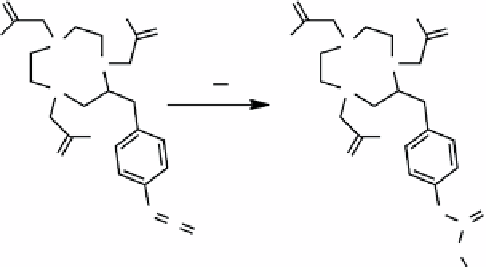
ScheMe 16.4
Reaction of
p
-NCS-Bn-NOTA, an empty chelate bearing an isothiocyanate group, with a generic amine results in
formation of a thiourea group and a strong covalent bond.
For MR imaging, the relaxometric properties were determined with the labelled MSNP system having an r
2
of 76.2 mM
-1
s
-1
versus the free SPIONs with a r
2
of 26.8 mM
-1
s
-1
. Three hours after injection, a hypointense signal at the tumour core was
observed, indicating that the particles had migrated to the tumour. Fluorescence measurements of
ex vivo
tissue sections
confirmed the delivery of the doxorubicin.
16.2.2
targeting applications of fluorescent-MrI bimodal agents
Hwang and co-workers added a targeting functionality to a multimodal nanoparticle system by using an aptamer targeted to nucleolin,
a cellular membrane protein highly expressed in cancer [7]. In this case, a silica-coated cobalt ferrite nanoparticle with an amine-
functionalised surface has been generated for the attachment of a rhodamine dye and the targeting aptamer. Additionally, a backbone-
modified chelate 2-(
p
-isothiocyanatobenzyl)-1,4,7-triazacyclonane-1,4,7-triacetic acid (p-NCS-Bn-NOTA) (Scheme 16.4) was
covalently attached to the amine-functionalised surface to allow for radioisotope labelling with
67
Ga. Utilising MRI and scintillation
images, they demonstrated successful targeting of the nanoparticle system to an
in vivo
mouse tumour model. In contrast, an NP
synthesised with a mutant aptamer rapidly cleared from the model and did not accumulate in the tumours.
Ex vivo
optical images of
the tumours confirmed the presence of the fluorescent NPs in the aptamer-targeted system and the absence in the mutant system.
A carbogenic nanocomposite has been described by Srivastava [26]. This nanocomposite was synthesised by a partial
thermal decomposition method using citric acid and l-lysine as organic precursors along with preformed SPIONs [27].
Through tuning the decomposition and growth of the organic layer, they demonstrate the ability to tune the fluorescent
response of the particle. The authors further report proof-of-principle MR and fluorescent images indicating that this non-
toxic system is effective as both a
T
2
and
T
1
agent. Fluorescent images of the
ex vivo
tissue sections confirm the results of the
MRI scans.
16.3
near-Infrared fluoreScence/MrI
The combination of NIR and MRI modalities and
in vivo
characterisation has been executed in two ways. The most straight-
forward method is to attach an organic NIR dye to a SPION (e.g., [28]). The most widely used commercially available
organic NIR dyes are based on a cyanine core (e.g., Cy5, Figure 16.4). The second approach is to combine SPIONs with a
materials-based NIR fluorescent species such as a quantum dot or multilayered gold/silica nanoparticles (e.g., [29]).