Chemistry Reference
In-Depth Information
Cl
-
N
+
N
O
CO
2
H
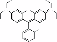
fIGure 16.2
Structure of rhodamine B.
NH
2
Br
Fe
3
O
4
O Fe
3
O
4
-MSN
Surface modi�ed dye-doped MSN
O
O
N
O
O
O
1)
H
3
C
n
O
O
2)
H
3
C
NH
2
n
DOX adsorption
DOX load Fe
3
O
4
-MSN
PEG coated Fe
3
O
4
-MSN
ScheMe 16.3
Functionalisation of MSNPs with SPIONs and DOX. Reproduced from Ref. [24] with permission from the American
Chemical Society.
Injection into a damaged optic nerve and injection into the eye of the mouse model resulted in increased infiltration of
activated microglia and macrophages but no peripheral toxicity in the subjects was observed. This result indicates that the
nanoparticles are nontoxic despite the activation of the immune system.
Particles were tracked using MRI for 14 days and confirmed in
ex vivo
tissue slices through optical imaging of the
fluorescent dye on the nanoparticles. The nanoparticles did not travel far from the injection site in either case and were not
observed in the peripheral tissue samples. These results show the promise of these types of agents being used as a platform
for sustained therapeutic delivery.
In a similar approach, Lee and co-workers have delivered chemotherapeutics to tumours in a mouse model using a meso-
porous silica nanoparticle (MSNP) [24]. They modified the MSNP surface with fluorescent dyes as well as SPIONs to pro-
vide both MR and optical visualisation (Scheme 16.3). SPIONs synthesised in organic solution were modified with
2-bromo-2-methylpropionic acid. These particles were mixed with MSNPs that possess free amine groups. Reaction bet-
ween the alkyl-bromide on the SPIONs and amine on the MSNPs provides a stable covalent bond. The porous nature of
silica allows loading of the system with a chemotherapeutic, in this case, doxorubicin (DOX) (Figure 16.3).


