Chemistry Reference
In-Depth Information
NH
2
NH
2
HO
HO
O
O
H
H
O
O
N
N
O
O
H
H
H
H
O
O
O
O
O
PGA
Glutaraldehdye
PLL
HO
NH
2
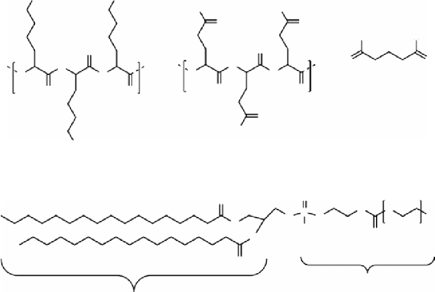
fIGure 16.5
Structures of polylysine (PLL), polyglutamic acid (PGA), and glutaraldehyde.
O
O
H
O
P
O
-
Na
+
O
OH
n
O
O
O
O
O
Hydrophilic
region
Hydrophobic
region
fIGure 16.6
Structure of DSPE-PEG 2000, n ~ 142 showing the hydrophobic and hydrophilic regions of the molecule that allow it to
form micelles.
Cell culture experiments with an external magnet suggest that the uptake by cells can be influenced by external magnetic
forces and confirmed that uptake can be tracked with fluorescence microscopy. The micelles were injected directly into
xenograft PC-3 tumours in athymic mice. The tumours were clearly observable and the overall signal intensity remained
constant for 24 h. No MR imaging was reported.
16.3.2
theranostic applications of nIr-MrI bimodal agents
Graphene is a strong NIR absorber and has potential use in photothermal therapy [37]. SPIONs can be grown from ferrous
chloride on reduced graphene oxide (RGO) by hydrothermal reaction [38]. The resulting nanocomposite can be further
functionalised with PEG-grafted poly(maleicanhydride-alt-1-octadecene) (C18PMH-PEG) for biocompatibility
(Scheme 16.6) [39]. The final product was found to be 50 nm in diameter with an r
2
of 108 mM
-1
s
-1
. Labelling the RGO-
Fe
3
O
4
-PEG particles with
125
I showed that they have a long blood circulation time. MRI and photoacoustic tomography
showed accumulation in tumours in mice. Irradiating the tumour with laser light (808 nm, 0.5 W/cm, 5 min) eradicated the
tumour after one day.
Mesoporous silica coatings on nanoparticles have been widely investigated for biomedical applications due to biocom-
patibility. The porous nature of this material allows drugs to be adsorbed for slow release
in vivo
. Ma et al. synthesised
oblong iron oxide particles coated with silica, followed by covalent attachment of gold nanorods (AuNRs) to make AuNR-
SPIONs (Figure 16.7) [40]. The AuNRs are known to be effective photothermal therapeutics. Further, modification of gold
nanoparticles is facile through the formation of a gold-thiol bond. Previous work has demonstrated the effectiveness of
this approach by using DNA modified with Gd(III) chelates and fluorescent dyes to modify gold nanoparticles for cell
labelling [10].
First, 200 nm hematite (Fe
2
O
3
) spindles were prepared by aging a solution containing Fe(ClO
4
)
3
.
6H
2
O, urea, and NaH
2
PO
4
in deionised water at 100 °C for 24 h. An amine-functionalised silica coating was added using TEOS and APTES, and the
particles were reduced to Fe
3
O
4
using a mixture of Ar and H
2
at high temperature. Separately, AuNRs were grown and mod-
ified with PEG and carboxylic acid disulfides to provide functionality. The AuNRs were covalently bound to the SPIONs
using the EDC/NHS coupling procedure. Doxorubicin was loaded by nonspecifically adsorbing into the silica shell pores.

