Chemistry Reference
In-Depth Information
many dual-modality SPECT/optical agents have also been developed. a tabulated summary of SPECT/optical agents is
provided in Table 15.2 and discussed in detail below.
15.3.1
small Molecule-Based agents
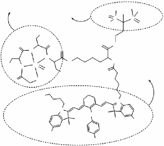
Possessing high binding affinity for hydroxyapatite (Ha) that is present in bone surfaces, bisphosphonates (BP) have been
extensively utilised to diagnose bone lesions and to treat breast cancer bone metastasis [73]. To target the microcalcification
in metastatic tumours, a BP-based dual-modality imaging agent (Pam-Tc-800) was developed [74]. In this probe, a lysine
residue was used as a linker to connect the three functional entities: 3-amino tetramethyl 1-hydroxy-propylidenebis-
phosphonate (Me-Pam) for Ha targeting [75], S-acetylmercaptoacetyltriserine (MaS
3
) for
99m
Tc-labelling [76], and the
NIRF dye 800CW (Figure 15.6a).
In vivo
studies of the agent in a breast cancer rat model showed high sensitivity detection
taBle 15.2
a tabulated summary of dual-modality spect/optical agents.
Radioisotope
Fluorophore
Targeting ligand
Target
References
99m
Tc
800CW
Bisphosphonate
Hydroxyapatite
[74]
99m
Tc
acridine orange
Bombesin peptide
gRPR
[77]
99m
Tc
acridine derivatives
Pyrazolyl-diamine ligands
DNa
[78]
99m
Tc
2-(4'-aminophenyl)benzothiazole
derivatives
2-(4'-aminophenyl)benzothiazole
derivatives
Cancer
(target unclear)
[79]
99m
Tc
Flavone
Flavone
β-amyloid
[80]
99m
Tc
Rhodamine 110
DEVD peptide
Caspase-3
[86]
111
In
800CW
c(RgDfK) peptide
Integrin α
v
β
3
[87, 88]
111
In
Cypate
c(RgDyK) peptide
Integrin α
v
β
3
[89]
111
In
IR-783
c(CgRRaggSC)NH
2
peptide
Il-11Rα
[95]
111
In
800CW
Trastuzumab
HER2
[103]
111
In
Cy5.5
Trastuzumab
HER2
[104]
111
In
Cy5.5/Cy7
Trastuzumab/Cetuximab
HER2/HER1
[105]
(a)
(b)
HO
OH
OH
99m
Tc-MAS
3
OH
Me-Pam
O
P
P
HO
O
OH
O
O
NH
O
O
N
N
HN
H
HO
Tc
N
O
O
OH
800 CW
S
O
NaO
3
S
+
N
N
O
-
SO
3
NaO
3
S
SO
3
Na
NIRF
SPECT/CT
FIgure 15.6
In vivo
SPECT/NIRF imaging of breast cancer microcalcification. (a) Chemical structure of the Pam-Tc-800 probe.
(b) NIRF and SPECT/CT images after administration of the probe in a breast cancer rat model. arrows indicate the tumour. adapted with
permission from Ref. [74]. (
See insert for colour representation of the figure.)
)