Chemistry Reference
In-Depth Information
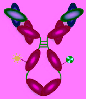
(a)
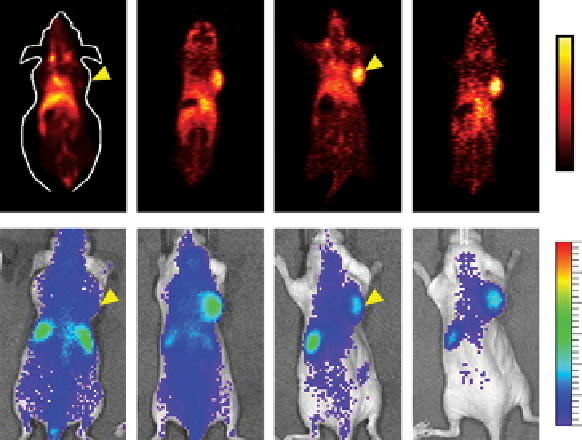
(b)
25 %ID/g
PET
0 %ID/g
4.0
3.0
NIRF
2.0
1.0
64
Cu-NOTA-Bev-800CW
4 h
24 h
48 h
72 h
(c)
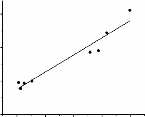
R
2
= 0.93
P<0.001
1.5×10
7
1.0×10
7
5.0×10
6
0.0
5
10 15
Tumour uptake (% ID/g)
20
25
FIgure 15.5
PET/NIRF imaging of VEgF. (a) Schematic representation of the dual-modality probe
64
Cu-NOTa-Bev-800CW. (b)
Serial
in vivo
PET/NIRF imaging of u87Mg tumour-bearing mice at 4, 24, 48 and 72 h post-injection of
64
Cu-NOTa-Bev-800CW.
arrowheads indicate the u87Mg tumours. (c) linear correlation of the
ex vivo
NIRF signal intensity with the percentage injected dose
per gram of tissue (%ID/g) values based on PET in all u87Mg tumour-bearing mice at 72 h post-injection [54]. (
See insert for colour
representation of the figure.)
)
In vivo
studies of the dual-modality agent revealed rapid, receptor-specific, and positive identification of the SlN in both the
NIRF and PET imaging modes, while maintaining little breakthrough to distal lymph nodes.
15.3
spect/optIcal agents
The source of SPECT images are gamma ray emissions [71, 72]. The first object that an emitted gamma photon encounters
after exiting the body is the collimator, which allows it to travel only along certain directions to reach the detector to ensure
that the signal position on the detector accurately represents the source of the gamma ray. Because of the use of collimators,
the sensitivity of SPECT is much lower than PET. The most common radioisotopes used for SPECT imaging include
99m
Tc
(t
1/2
: 6.0 h),
111
In (t
1/2
: 2.8 days), and radioiodine (e.g.
131
I, t
1/2
: 8.0 days). Similar to the abovementioned PET/optical agents,