Chemistry Reference
In-Depth Information
lymphatic UCL imaging using NaYF
4
:Yb,Er and NaYF
4
:Yb,Tm nanoparticles [76]. Li and co-workers further applied
amphiphilic LaF
3
:Yb,Tm nanoparticles to image lymph nodes using NIR upconversion emission as the output signal
(Figure 13.7) [77]. Liu and co-workers employed three different kinds of Ln-UCNPs (NaY
0.78
Yb
0.2
Er
0.02
F
4
, NaY
0.69
Yb
0.3
Er
0.01
F
4
,
NaY
0.78
Yb
0.2
Tm
0.02
F
4
) with multicolour emissions for UCL imaging of lymph nodes at different positions [52]. These results
demonstrate that the lymph nodes can be easily imaged by upconversion luminescence. Benefitting from the high sensitivity
and signal-to-noise ratio, this imaging technique has potential in the luminescent imaging-guided surgery to remove the
lymph nodes infected by cancers.
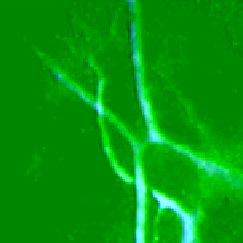
13.4.4.4 Vascular Imaging
vascellum is one of the most important organs in animals to transport blood around the body.
vascular imaging can provide information about the shape, position, spacing, permeability, for example. Ln-UCNPs can also
be used for vascular imaging after proper surface modification to prolong their existence in the blood. Hilderbrand and
co-workers first used PEg-coated Y
2
o
3
:Yb,Er nanoparticles for blood-pool UCL imaging via intravenous injection of the
Ln-UCNPs. A NIR-emitting carbocyanine fluorophor was also used for co-localisation imaging (Figure 13.8) [78]. Zhang
and co-workers also applied both NaYF
4
:Yb,Er@Sio
2
nanoparticles and NaYF
4
:Yb,Er@Sio
2
-labelled cells for the UCL
imaging of mouse ear blood vessels [79]. Furthermore, van veggel et al. employed
in vivo
two-photon upconversion wide
field microscopy to image brain blood vessels of a mouse after skull thinning [80].
13.4.4.5 Cell Tracking
Another imaging application is to track cells in animals. Because some kinds of cells can be
easily labelled by Ln-UCNPs as described in section 13.4.3.1, the upconversion luminescence from those labelled cells can
help to track the motion and accumulation of these cells.
Zhang and co-workers first used NaYF
4
:Yb,Er@Sio
2
nanoparticles to label myoblast cells, which were introduced to a
mouse model with a cryo-injured hind limb [79]. The upconversion emission signals can be detected inside the limb of the
Brighteld
UCL
Overlay
1150
2862
ALNs
ALNs
3,4
3,4
4575
2
2
1
1
6287
Inject site
Inject site
0.5 cm
0.5 cm
0.5 cm
8000
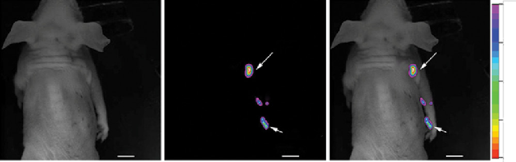
FIgure 13.7
In vivo
lymphatic drainage upconversion imaging at 800 nm to show four different draining lymph basins (1, 2, 3, 4)
along the right antebrachium of the nude mouse. Reprinted with permission from Ref. [77]. Copyright 2011 Elsevier Ltd. (
See insert for
colour representation of the figure.)
)
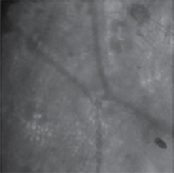
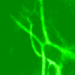
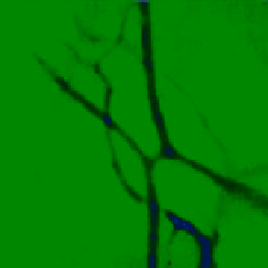
(a)
(b)
(c)
(d)
FIgure 13.8
optical imaging of blood vessels in the mouse ear following tail vein injection of the nanoparticles (10 mg); (a) blood
vessels imaged with a blue light filter, (b) upconversion image with excitation at 980 nm, (c) fluorescence image of the carbocyanine dye
with excitation at 737 nm, (d) merged image of the upconversion and fluorescence signals. Reprinted with permission from Ref. [78].
Copyright 2009 The Royal Society of Chemistry. (
See insert for colour representation of the figure.)
)