Chemistry Reference
In-Depth Information
mouse, demonstrating that upconversion imaging can be performed as a noninvasive technique to track cells
in vivo
. Liu et al.
further applied NaY
0.78
Yb
0.2
Er
0.02
F
4
, NaY
0.69
Yb
0.3
Er
0.01
F
4
, and NaY
0.78
Yb
0.2
Tm
0.02
F
4
with different upconversion emission
properties to label cancer cells [52]. Seven days after subcutaneous injection of labelled cells, they also found the tumour at
the location of injection, which agreed with the UCL cell-tracking imaging. This result indicates that the labelling protocol
will not destroy the bioactivity of cancer cells. Further experiments are needed to track these cells during a long period to
investigate the metastasis or other biological processes involving living cells.
13.4.4.6 Tumour Targeting
In vivo
Tumour-targeting techniques have attracted increasing attention, because this is a
key point in tumour diagnosis and therapy. Ln-UCNPs have been successfully applied for tumour target imaging in a few
cases, based on the ligand-acceptor or antigen-antibody interactions. However, the tumour-targeting properties originate
from the active species linked to the Ln-UCNPs, rather than the nanoparticles themselves. Similar to the case for cell
imaging, folic acid and peptides are still two kinds of specific active groups for tumour targeting.
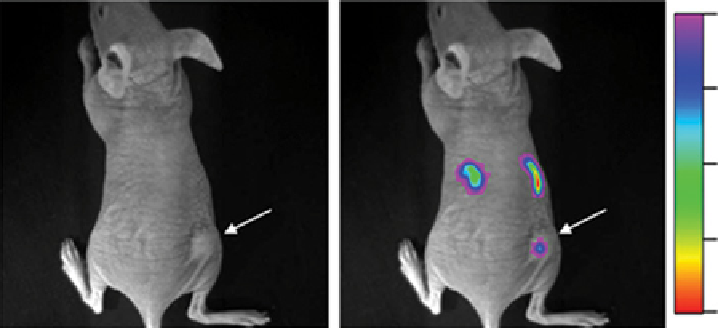
on the basis of the high affinity between FA and FR, Li and co-workers reported the first example of imaging
FR-overexpressed tumours in small animals model (Figure 13.9) [67]. FA-modified Ln-UCNPs were intravenously injected
into a nude mouse with HeLa tumour-bearing athymic. Upconversion emission signals at 600-700 nm were collected from
the position of the tumour. As a control experiment, amine-functionalised Ln-UCNPs were intravenously injected into the
nude mouse via the same tumour, but no significant upconversion signals could be detected. A blocking dose of FA (10 mg/
kg) was also introduced to the mouse to block the target effect.
Li and co-workers also used arginine-glycine-asparatic acid (RgD) peptide, which has a high affinity for the α
v
β
3
integrin
receptor, to target tumour tissues [3]. Cyclopeptide c(RgDFK) was conjugated to NaYF
4
:Yb,Er,Tm nanoparticles with the
(a)
Brighteld
Merge
95.00
121.25
147.50
173.75
199.99
(b)
95.00
116.56
138.13
159.69
181.25
FIgure 13.9
In vivo
UCL imaging of subcutaneous HeLa tumour-bearing athymic nude mice (right hind leg, pointed by white arrows)
after intravenous injection of UCNPs-NH
2
(a) and UCNPs-FA (b). All images were acquired under the same instrumental conditions
(power density = 120 mW/cm
2
on the surface of mouse). Reprinted with permission from Ref. [67]. Copyright 2009 Elsevier Ltd.