Chemistry Reference
In-Depth Information
Recently, Yan et al. used a similar system to evaluate the metabolism of Ln-UCNPs through the digestive system. [74].
NaYF
4
:Yb,Tm can be eaten by
C. elegans
and found in the gut from the pharynx to the anus. After being fed with
Escherichia
coli
,
C. elegans
will excrete these Ln-UCNPs, and no upconversion signals can be found.
13.4.4.2 Animal Whole-Body Imaging
Apart from
C. elegans
, mice and rats are the most commonly used small animal
models upon which upconversion imaging has been performed. Zhang and co-workers reported
in vivo
upconversion imaging
near the body surface and deep in the body of rats [66]. The emission at 800 nm from PEI-coated NaYF
4
:Yb,Tm nanoparti-
cles can be used as a detection signal. Following this, many research groups have used Ln-UCNPs as luminescent labels for
the
in vivo
bioimaging in animals, and the nude mouse is the common animal model because of the minimum scattering
effect. However, upconversion imaging of mice with fur can also be achieved. For example, Prasad's group also applied
NaYF
4
:Yb,Tm nanoparticles for
in vivo
imaging [75]. They proved that this upconversion imaging provides deeper light
penetration with almost no autofluorescence.
Liu and co-workers reported a penetration depth of 0.8 cm for upconversion imaging, which was evaluated in packing
pork using NaYF
4
:Yb,Tm as probes [59]. Li and co-workers applied NaLuF
4
-based UCNPs with higher upconversion
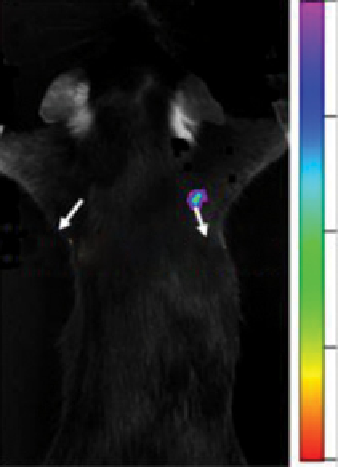
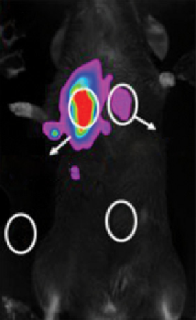
efficiency for imaging to achieve larger penetration depth (Figure 13.6) [4]. Black mice, in which the absorption of excita-
tion and emission are larger than in nude mice, were employed as the animal model. By injecting subcutaneously with
NaLuF
4
:Yb,Tm nanoparticles, upconversion signals can be collected from both the front and back sides of the mouse, indi-
cating the successful penetration of the whole mouse with a penetration depth of ~ 2 cm.
Furthermore, Li and co-workers applied NaLuF
4
:Yb,Tm nanoparticles obtained by solvothermal methods for the upcon-
version imaging of a rabbit [60]. In this large animal model, upconversion imaging signals can also be collected with excel-
lent signal-to-noise ratio.
As described above, upconversion imaging can be achieved for mice and rabbits for whole body imaging. However,
because no target species were linked to the Ln-UCNPs in these examples, the imaging signals generally locate in the lung,
liver, and spleen based on the biodistribution of Ln-UCNPs (which will be discussed in section 13.5).
13.4.4.3 Lymphatic Imaging
The lymph node is an important organ of the immune system in mammals; it has the ability
to trap foreign particles and give the corresponding signals to the immune system. Lymph nodes are the most common place
for cancers especially to be metastasised. Therefore, the imaging and position of lymph nodes have essential significance in
clinical and biological applications. Because of the ability of lymph nodes to trap foreign particles, Ln-UCNPs can accumu-
late in lymph nodes easily after injection in the paw of a mouse. Kobayashi and co-workers first obtained two-colour
(a)
(b)
1000.0
4875.0
1ʹ
1
Cit-Y1-Tm
8750.0
Cit-Lu6-Tm
Cit-Y1-Tm
Cit-Lu6-Tm
2
3
12625.0
16500.0
FIgure 13.6
Upconversion imaging of a black mouse by subcutaneous injection of NaLuF
4
and NaYF
4
-based UCNPs, (a) detection
from the side of chest, (b) from the side of back.
λ
ex
= 980 nm,
λ
em
= 800 nm. Reprinted with permission from Ref. [4]. Copyright 2011
American Chemical Society.