Chemistry Reference
In-Depth Information
are located in the microemulsions, while the silica shell forms at the interface between the aqueous solution and organic
solvent (usually cyclohexane). The silica shells obtained in microemulsions are usually uniform, but the size of the entire
Ln-UCNPs@Sio
2
is limited by the size of the droplets. When the size of Ln-UCNPs@Sio
2
is larger than the droplets, the
hydrolysis of TEoS cannot be well controlled to occur at the interface, resulting in non-uniform products.
After silica coating, functional groups such as -NH
2
can be directly induced to the Ln-UCNPs by applying silica sources
containing -NH
2
groups (APS). Researchers have also developed a method of constructing mesoporous silica shells outside
Ln-UCNPs, which enables a larger loading amount of the structure.
In addition to the post-surface modification method, there are also some reports on the direct synthesis of surface
functional Ln-UCNPs. In these methods, functional groups are introduced to the Ln-UCNPs during the synthesis procedure,
usually acting as moieties of the capping ligands.
13.4
In VIVo
ImagIng aPPLIcatIonS
13.4.1
Setup of Imaging Instruments
Because most commercial microscopes and cameras are based on the detection of visible emissions, imaging instruments
should be designed and modified to obtain upconversion imaging.
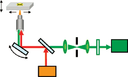
For microscopes, upconversion has luminescent characteristics similar to two-photon luminescence. Therefore, the
instrument can be set up on a similar microscope. Li and co-workers chose a commercial inverted microscope (olympus
IX81) with a confocal scanning unit (Fv1000, olympus, Japan) as the base to construct the imaging instrument
(Scheme 13.3a) [2]. A continuous-wave laser emitting at 980 nm was introduced as the excitation source. The laser beam was
(a)
Specimen
z scanning stage
Objective lens
Reverse excitation
dichroic mirror
Detector
Galvanometer
mirrors
Confocal
pinhole
Filter
CW Laser at 980 nm
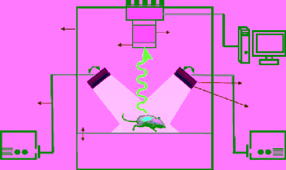
(b)
EMCCD
Computer
Light-tight
housing
Lens
Emission
�lter
Beam
expander
980 nm
fuber optics
Lens
The sample table
CW laser
CW Laser
Scheme 13.3
Schematic illustration of the instruments for cell imaging (a) and animal imaging (b).