Chemistry Reference
In-Depth Information
(a)
(b)
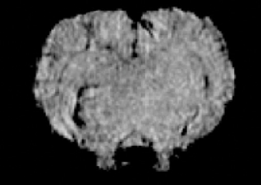
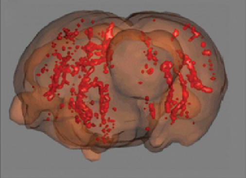
FIgure 9.12
(a) selected image taken from T
2
*
weighted 3D dataset of rat brain. (b) 3D reconstruction of the accumulation of contrast
agent. The images reveal that magnetic nanoparticles functionalised with sLe
x
enable detection of lesions in models of stroke. Reproduced
with permission from Ref. [154]. (
See insert for colour representation of the figure.)
)
they are weaker than other biological interactions so a multivalent effect is usually needed to measure or observe them.
Nanoparticles are thus a perfect platform to display carbohydrate motifs with the multivalent presentation needed to study these
interactions. In this report, iron oxide nanoparticles (Fe
3
O
4
) were prepared following a co-precipitation protocol starting from
iron(III) chloride and iron(II) sulphate salts and without the presence of any ligand. Naked Fe
3
O
4
nanoparticles were subsequently
coated with a polyvinylpyrrolidone (PVP) polymer and a stable silica shell (using TeOs as the siO
2
precursor). After some
manipulation, simple carbohydrates were coupled to the nanoparticles using standard peptidic coupling conditions (mannose,
galactose, fucose, or sialic acid) with the APTes-functionalised nanoparticles, or click chemistry was used with the azide-func-
tionalised ones (N-acetylglucosamine). The group acquired T
2
-weighed MRI images and calculated the variation in T
2
for each
nanoparticle. Their results showed that each kind of cells has a different carbohydrate-binding characteristic that could be used
for differentiation, and more importantly, that the carbohydrate-binding pattern changes from healthy to tumoural cells.
Van Kasteren et al. presented research using carbohydrates as targeting molecules [154]. In this case, they wanted to dem-
onstrate that magnetic nanoparticles functionalised with complex carbohydrate structures could be used to image activated
brain endothelium. This activation is associated with the inflammation process that takes place in diseases such as multiple
sclerosis, ischemic stroke, and HIV-related dementia, and cannot be visualised by conventional imaging techniques. To
image this process, e-/P-selectins (CD62e/CD62P) were chosen that are endothelial markers in acute inflammation. These
molecules are carbohydrate-binding transmembrane proteins whose known ligand is sialyl Lewis
x
, so a chemical strategy
was designed to form iron oxide nanoparticles with these ligands on their surface. The coupling of the ligand to the nanopar-
ticle was achieved via the reaction of an imidoester in the carbohydrate to an amine group on the nanoparticle. The final
results obtained by MRI (Figure 9.12) using these probes
in vivo
with different animal models of brain diseases combined
with hystochemical corroboration, confirmed that these nanoparticulate probes were able to target the activated endothelia
when the contrast agent was administrated via the tail vein.
A different and simpler strategy was followed by Nieman and co-workers to study the migration of stem cells in brain
[160]. Commercial iron oxide-based nanoparticles encapsulated inside a polymer microbead were purchased and used
without further modification. In this report, instead of trying to design a complex strategy to target certain cells, the probes
were delivered directly to their target through a stereotactic injection. The intention was to follow
in vivo
the migration of
stem cells from their niche in the subventricular zone to the olfactory bulb in a physiological process to maintain the homeo-
stasis of olfactory neurons using MRI (Figure 9.13). They demonstrated that upon labelling of a certain population of cells
(neural stem cells in this case), these can then be tracked non-invasively
in vivo
along several days with a precision that
allows the calculation of speeds of migration and allows the anatomic placement of the cells in their destination.
Hadjipanayis et al. have prepared and functionalised magnetic nanoparticles to be used as MRI probes for the detection
and treatment of glioblastoma multiforme [161]. The preparation of the magnetic nanoparticle probes was from commercial
iron oxide nanoparticles coated with an amphiphilic triblock copolymer. This copolymer included PeG moieties to minimise
non-specific interactions and carboxylic groups that allow them to continue the functionalisation of the nanoparticles. using
amide-coupling conditions (eDC/sulfo-NHs), a specific antibody anti-epidermal growth factor receptor (eGFR) deletion