Chemistry Reference
In-Depth Information
fluorescence imaging). Here, we will only give a few examples of purely T
1
MRI agents; multifunctional nanomaterials
will be discussed in Chapter 16.
silica and gold nanoparticles have been used as platforms onto which the different chelates can be coupled. silica
nanoparticles present the advantage that they can be synthesised as mesoporous materials comprising channels with diame-
ters tunable between 2 and 10 nm. This feature allows their use either as platforms onto which molecules can be attached or
as encapsulating materials containing active species in their channels. The work by Weili and Lin [58, 59] demonstrated that
gadolinium chelates can be bound both to the surface or the channels of silica nanoparticles. In reference [58] 37 nm silica
(siO
2
) nanoparticles incorporating gadolinium chelates in their surface were prepared following a two-step one-pot reverse
microemulsion protocol. Alternatively, in reference [59], mesoporous siO
2
nanoparticles were prepared following a surfac-
tant template, base-catalysed condensation procedure. Cetyltrimethylammonium bromide (CTAB) was used as the surfac-
tant in water, and TeOs was used as siO
2
precursor (Figure 9.4a, b). The polymerisation was catalysed by NaOH and carried
out for 2 hours at 80°C. According to their data, the Gd chelate is mainly attached to the channels in the structure of the
nanoparticles. This channel location improves the performance as T
1
contrast agents because it seems to present highly effi-
cient accessibility of the magnetic centres to available water molecules.
(a)
(b)
-
O
O
N
O
N
O
O
O
Si
Gd
=
O
O
N
O
O
O
OH
2
OH
2
2 μm
Gd-Si-DTTA
(c)
Au
Au
CuAAC
“click”
H
H
O
N
O
O
O
O
O
O
=
Au
= Gold nanoparticle 13 nm
Gd
O
N
N
N
N
T
=dT-N
3
= 3'SH-T9
T
TT
T
TT
T
TT
T
TT
T
TT-
Cy3
-5'
(d)
1.4 nm
20-80 nm
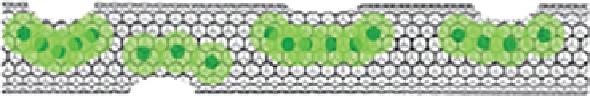
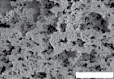
FIgure 9.4
(a) seM image of mesoporous silica nanospheres showing the formation of monodisperse, water-dispersible nanoparti-
cles. (b) scheme showing the Gd-si-DTTA complexes residing in hexagonally ordered nanochannels of 2.4 nm in diameter. Reproduced
with permission from Ref. [59]. (c) scheme of the DNA-GdIII@AuNP conjugates prepared in Ref. [63]. (d) Representative illustration of
a gadonanotube (GNT). Clusters of internally loaded Gd
3+
ions are located at defect sites along the nanocapsule sidewalls. Reproduced
with permission from Ref. [65].