Biomedical Engineering Reference
In-Depth Information
implant than preculturing them for
days. For
the other groups, no signifi cant differences
were observed. Furthermore, no signifi cant
differences were observed between implants
cultured under various conditions, including
static and fl ow perfusion. However, it seems
that preculturing cells for
4
day under fl ow per-
fusion enhanced bone formation more than
preculture under static conditions. The results
of this study are consistent with those of a pre-
vious study demonstrating that bone formation
in an orthotopic site was more effectively
induced by a short preculture of osteogenic
cells after seeding in titanium fi ber mesh.
However, these results provide only weak evi-
dence that fl ow perfusion in the present form
has the potential to increase bone formation in
an orthotopic site. Comprehensive testing and
verifi cation in a modifi ed experimental setting
are needed before fl ow perfusion can be
assumed to increase bone formation.
1
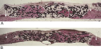
Figure 5.4.
Light micrographs of implants loaded or not
loaded, with rat bone marrow cells after implantation for 30
days. Arrows indicate the edges of the bone defects. A.
Unloaded implants. B. Cell-loaded implants. Some of the tita-
nium implants showed bone formation, morphologically char-
acterized by the occurrence of osteocytes embedded in a
mineralized matrix (1.6
×
magnification). Bonelike tissue was
distributed uniformly in all titanium-rat bone marrow implants.
Union of skull bone with newly formed bone in the implant
was also observed at one side of the implant (1.6
×
magnification).
cluded that inoculating titanium fi ber mesh
with bone marrow cells improves the bone-
healing capacity of this material.
Earlier in vitro studies had shown that
dynamic culturing, especially the fl ow-perfu-
sion system, enhanced the osteogenic differen-
tiation and growth of cells inside the meshes.
In vivo studies had not yet investigated the
effect of this osteogenic improvement on the
fi nal osteoinductive properties of the titanium
mesh constructs. Therefore, cell-seeded meshes
were precultured for
5.6 Growth-Factor-Based
Approach: Titanium
Fiber Mesh
Numerous in vivo experiments have been per-
formed to evaluate the effect of growth factor-
coated titanium fi ber mesh on bone formation.
The osteoinductive properties of porous tita-
nium fi ber mesh with a calcium phosphate
coating loaded with rhBMP-
days under
static conditions or with the fl ow-perfusion
system [
1
,
4
, and
8
were subcutane-
ously placed in Wistar rats and implanted for
2
]. After culture, cell-loaded implants
were placed in an
32
3
8
-mm cranial defect and
to
days. Histological analysis demonstrated
the induction of ectopic cartilage and bone
formation by
40
retrieved after
days of implantation
for both histological and histomorphometrical
examinations. After
7
and
30
9
days, cartilage was seen together with trabecu-
lar bone. At
5
and
7
days, respectively. At
days of implantation,
bone formation was absent in all groups.
Further, the fi ber mesh porosity was fi lled with
fi brous tissue containing capillaries. After
7
days, bone formation had
increased and was characterized by the pres-
ence of trabecular bone and bone marrow-like
tissue. At
20
30
days of implantation, most implants showed
bone formation, except for one implant precul-
tured for
days, more lamellar bone and
hematopoietic bone marrow-like tissue were
present. Thus, calcium phosphate-coated tita-
nium fi ber mesh containing rhBMP-
40
days under fl ow-perfusion condi-
tions and one implant precultured for
4
days
under static conditions. Both blood vessels and
bone marrow were observed. The results of the
histomorphometrical measurement showed
that preculturing cells for
8
can
induce ectopic endochondral-like bone forma-
tion in a rat model over short implantation
periods [
2
].
In another study, rhTGF-
43
,
44
day in the fl ow-
perfusion system produced a signifi cantly
higher percentage of bone present in the
1
β
1
-loaded titanium
fi ber meshes were implanted in a New Zealand
white rabbit noncritical-size cranial-defect