Biomedical Engineering Reference
In-Depth Information
occurred between days
. SEM and
calcium measurements showed an increase in
calcium from days
4
and
8
demonstrated that bone had formed in all day-
1
and day-
implants, and that the bonelike
tissue was present uniformly through the
meshes. The bony tissue was morphologically
characterized by the occurrence of osteocytes
embedded in a mineralized matrix with a layer
of osteoid and osteoblasts at the surface. In the
day-
4
, with only a
small, although signifi cant, increase between
days
1
through
4
4
and
8
(Fig.
5
.
3
). Histological analysis
implants calcium phosphate had depos-
ited only in the titanium fi ber mesh. Calcium
measurements of the implants revealed that
calcifi cation in day-
8
1
implants was signifi cantly
higher than in day-
implants. No
signifi cant difference in calcium content was
observed between day-
4
and day-
8
implants.
On the basis of these results, we concluded that
bone formation was enhanced by a short culture
time of osteogenic cells after seeding in tita-
nium fi ber mesh and that dynamic cell seeding
is probably more effective than static cell
seeding.
Although the cell-loaded meshes demon-
strated osteoinductive properties in a subcuta-
neous model, it was important to evaluate the
bone regenerative properties of cell-loaded
titanium fi ber meshes in a bony environment
[
4
and day-
8
]. Therefore, meshes with cells were subcul-
tured for
37
day under standard conditions.
Cell-loaded implants and controls then were
placed in an
1
days
of implantation, mineralized-like matrix depo-
sition and blood cells were observed inside the
mesh porosity of both groups. In addition to
blood cells, blood vessels were visible in two
out of six of the cell-loaded specimens. After
8
-mm cranial defect. After
3
15
days of implantation, only one out of six control
implants showed bone formation inside the
implant porosity, but bone was present uni-
formly throughout all cell-loaded meshes.
Blood vessels and bone marrow were also
observed. Only two cell-loaded implants
showed union at the cranial defect perimeter.
After
days of implantation, all cell-loaded
implants showed bone formation inside the
mesh, but in the control group only four of six
implants had produced new bone (Fig.
30
).
Bone marrow and bone union at the bone defect
borders were found only in fi ve out of eight of
the cell-loaded implants. The histomorpho-
metrical evaluation found that no bone tissue
was present in either implant group after
5
.
4
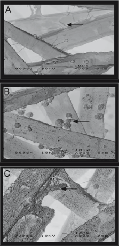
Figure 5.3.
Scanning electron micrographs show that (A)
after 1 day of culture, fibers were covered with layers of well-
spread osteogenic cells (arrow). (B) After 4 days of culture, the
deposition of a calcified matrix, characterized by the occur-
rence of globular accretions (arrow), could already be recog-
nized. (C) After 8 days of culture, calcification appeared to
increase, and large and small globular accretions as well as
collagen bundles (arrow) covered the fibers almost
completely.
3
days
of implantation, and that after
days,
signifi cantly more bone was present in cell-
loaded implants than in unseeded control
implants. On the basis of these results, we con-
15
and
30