Biomedical Engineering Reference
In-Depth Information
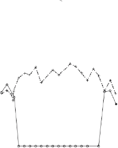
Figure 8.3.
Bone formation rates (A)
percentage trabecular bone area and (B)
mineral appositional rate, measured in
the region of interest, reached a
maximum 4 weeks after the end of dis-
traction. Data adapted from Samchukov
et al, 2001 [101].
A. Percentage Trabecular Area
100
80
Control
0 Week
2 Weeks
4 Weeks
6 Weeks
8 Weeks
60
40
20
0
0
5
10
15
20
ROI
B. Mineral Apposition Rate
3
2.5
Control
0 Week
2 Weeks
4 Weeks
6 Weeks
8 Weeks
2
1.5
1
0.5
0
0
5
10
ROI
15
20
The effect of motion on distraction regenera-
tion relies on two mechanisms: formation of
fi brovascular matrix, and growth and condensa-
tion of hydroxyapatite crystals. We propose the
use of a stress-strain diagram (Fig.
all threee phases (cartilage, fi brous cartilage,
and osscous tissue) can be developed. Molecular
studies have shown that during the early stage of
healing, motion inhibits mesenchymal cell dif-
ferentiation into osteoblasts by increasing
expression of the
ihh
gene, which regulates chon-
drocyte maturation during fetal and early post-
natal skeletogenesis [
) to predict
differential bone phases analogous to the general
chemistry phase diagram that is used to explain
the relationship between ice, water, and water
vapor. This diagram is a modifi cation of Carter's
previous work [
8
.
5
]. Collagen, ECM proteins
and hydroxyapatite crystals are associated with
the osseous phase. Cytokines such as IGF-
82
] and is based on histomorpho-
metric observations. The three phases of bone
formation (endochondral) are the fi brous, carti-
lage, and osseous phases. Neither the fi brous nor
the cartilage phase contains hydroxyapatite,
which means that Ca
2
+
and PO
4
3
−
ions in the
tissue do not form apatite crystals. High tensile
stress and strain cause fi brous tissue formation,
while high compression produces osseous tissue
(tissue mineralized with hydroxyapatite). In the
area between low tensile and low compression,
16
1
,
TGF-
, which can modulate
mineralization, are up-regulated in cells associ-
ated with distracted tissues [
β
, BMP-
2
, and BMP-
4
]. The formation
of these phases is controlled by the products of
the cells (e.g., collagen, and ECM proteins) and
physical-chemical reactions among the secreted
cell products and calcium and phosphate ions.
The stress/strain history of the tissues can affect
both the cell products and the subsequent physi-
cal-chemical interactions.
104