Biomedical Engineering Reference
In-Depth Information
A.
B.
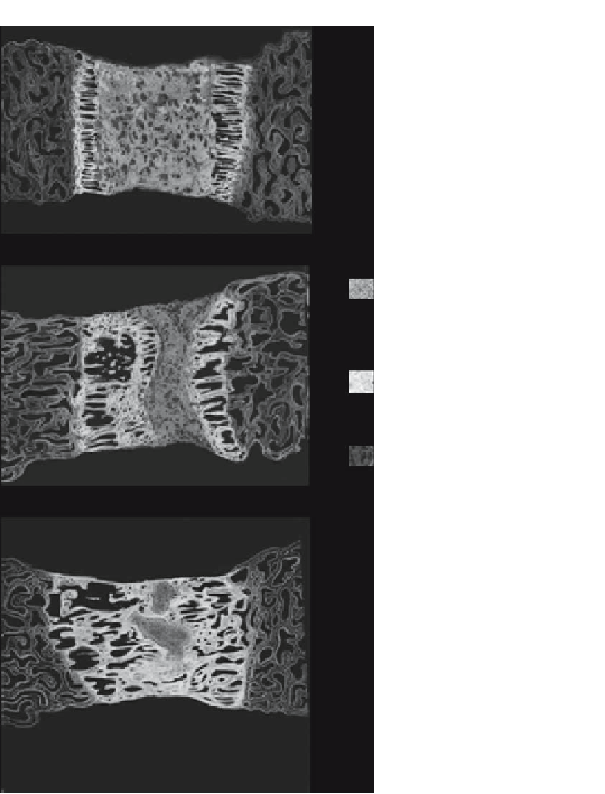
fibrovascular interzone tissues
new bone
old bone
C.
Figure 8.4.
Schematics showing histomorphology of distraction regeneration at week 0 (A), week 2 (B), and week 4 (C) after
distraction of rat mandible. The proportion of old bone, new bone, and fibrovascular interzone tissues varied at different stages.
Diagram was adapted from the original histological findings of Samchukov et al [101].