Biomedical Engineering Reference
In-Depth Information
their effects on bone formation, because this
technique produces a large volume of new bone
in a controlled fashion [
22
2
1.8
1.6
1.4
1.2
1
0.8
0.6
0.4
0.2
0
]. The technique
of distraction osteogenesis has been used in the
practice of orthopedics and oral maxillofacial
surgery. The procedure, following osteotomy,
includes a latency period of up to
25
,
98
Stretch
Ratio
days, a dis-
traction period during which the osteotomized
gap is lengthened by
6
0
.
50
to
0
.
75
mm per day
for
8
weeks. These procedures were designed on
the basis of the compliance or stiffness of the
ECM and result in increasing the length of bone
by
14
days, and a consolidation period of
0
5
10
15
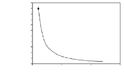
Figure 8.2.
Stretch ratio, defined as deformation divided by
the original length of the distracted gap, during distraction
osteogenesis. The stretch ratio decreases as the distraction
period increases.
cm, depending on the duration of
distraction.
The latency period is the period from bone
division to the onset of traction and represents
the time required for reparative callus forma-
tion. Prolonging the latency period of bone
healing may prevent distraction. If the bone
matures to a point at which mineralization is
signifi cant, the distraction process will frac-
ture bone instead of inducing growth. As bone
matures, it accumulates hydroxyapatite and
becomes brittle, giving it very little deforma-
tion range. Thus, the correct length of the
latency period should be determined before
proceeding to the distraction stage. Inadequate
scheduling of each phase may result in relapse
and failure to lengthen [
1
to
10
tion of the compression or the shear strain,
corresponds well with the direction of fi brous
tissue formation.
The consolidation stage represents the time
at which large amounts of hydroxyapatite are
deposited in bone. The rate of bone formation
appears to reach a maximum
2
to
4
weeks after
the completion of distraction (Fig.
). At the
beginning of consolidation (end of distraction),
the tissue is fi lled with fi brovascular tissue
comprising
8
.
3
% of the total regenera-
tion area and and organized as parallel colla-
gen bundles with interspersed vascular
channels. Only
70
% to
93
].
Formation of soft callus is the key to success-
ful distraction and vascularization. The soft
callus includes collagen and progenitor cells.
The distraction appears to be related to the
movement of fl exible, threadlike, long-chain
molecules of collagen. Long collagen fi bers
along the direction of stretch have been
reported in the distraction gap. When collagen
undergoes changes in confi guration, internal
cohesive force (stress) is developed. This
internal stress is then transferred into the
cells, where collagen transcription rates are
increased. Like a manufacturing plant, the cells
produce large amounts of ECM until the gap is
fi lled. It has been shown that the increased
products in the distraction stage consist of
primary fi brous and vascular tissues; little
hydroxyapatite has been found. The strain
decreases every day during distraction from
infi nite strain (day
1
,
111
% of the regenerated
tissue consists of bony trabeculae; the remain-
ing
2
% to
5
4
% to
27
% is marrow space (Fig.
8
.
4
A). At
2
weeks of consolidation, new bone formation
occupies up to
30
% of the distracted region
(Fig.
8
.
4
B). At
4
and
6
weeks, new bone occupies
40
% of the area, with a small fi brous
interzone remaining (Fig.
% to
45
weeks, the
regenerated area is fi lled with trabecular bone
and lacks a fi brous interzone. The trabeculae
increase in both length and thickness during
the time of consolidation and are oriented par-
allel to the direction of distraction. No tensile
strain greater than
8
.
4
C). At
8
should be applied to the
tissues during the consolidation stage, because
excessive distraction inhibits crystal growth.
However, Richards [
0
.
1
] has shown that adding
small compressive strains (strains less than
0
98
) might provide additional stimu-
lation to cells and produce more bone than
would be produced without any additional
forces. In addition, pressure at the local site
may produce a consolidation effect on the for-
mation of hydroxyapatite crystals and hydroxy-
apatite-collagen complex.
.
003
;
3000
µε
1
) to less than
0
.
1
strain by
day
). The strain level appears much
higher than the levels that Frost and other
researchers reported to occur during bone
remodeling [
10
(Fig.
8
.
2
]. In addition, the direc-
tion of the tensile strain, rather than the direc-
38
,
84
,
100