Biomedical Engineering Reference
In-Depth Information
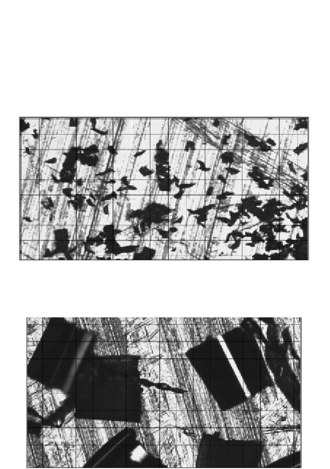
comparison purposes after impacts were carried out. Microscopic images with
these particles can be seen in Figure 50 and Figure 51. Specifications for the
microscope and camera are listed in Table 5. Each type of particle was then
impacted onto each type of substrate, with the numbering specified in Table 6.
Three techniques to investigate analyze effects of impacts were employed:
chemical composition analysis, hardness testing, and microscopic imaging.
Additionally, high-speed imaging is used to further gain insight into the impact
events.
Reproduced with permission. Copyright retained by Inderscience Publishers.
Figure 50. Fine Aluminum Particles with 100 µm Grid.
Reproduced with permission. Copyright retained by Inderscience Publishers.
Figure 51. Coarse Aluminum Particles with 100 µm Grid.