Gram-positive bacteria appear bluish-purple in the Gram stain, while Gram-Negative Bacteria are red. The crystal violet-iodine complex formed during Gram-staining is retained by the cell wall of Gram-positive bacteria.
Gram-positive eubacteria (bacteria) are found in several taxonomic groups. Typical representatives are both endospore-forming and nonsporing rods of the genera Bacillus, Clostridium, Arthrobacter, Lactobacillus, Mycobacterium, Nocardia and Streptomyces. A few archaebacteria (Archaea), such as Methanobacterium formicicum, also stain Gram-positive. The Gram reaction depends on both the structural formats and the chemical composition of the cell envelope (see Gram-Negative Bacteria). Gram-positive bacteria have a broader range of cell-wall types than do Gram-negative bacteria (1).
1. Type I Gram-positive cell wall
This type of cell wall consists of a thick peptidoglycan layer mixed with teichoic acids. Peptidoglycan (murein) is a polymer of glycan strands of N-acetyl-D-glucosamine and N-acetylmuramic acid linked in alternating sequence by b-1,4 linkages. The glycan strands are cross-linked by peptide chains of varying complexity, which are bound in amide links to the carboxyl group of the lactic acid residue of muramic acid (Fig. 1a). In Gram-positive bacteria, the amino acid composition of the peptides cross-linking the glycan strands (interpeptide bridges; Fig. 1a) vary considerably. In these interpeptide bridges, D-amino acids dominate, while aromatic, branched-chain, and sulfur-containing amino acids, plus histidine, arginine, and proline, are absent. In the tetrapeptide of Gram-positive bacteria, diaminopimelic acid can be replaced by lysine.
Figure 1. Schematic structure of polymers in cell walls of Gram-positive bacteria. A. Peptidoglycan with an polyglycine interpeptide bridge, [Gly]5 in Staphylococcus aureus. B. Teichoic acid of the ribitol-phosphate type. C. Lipoprotein with fatty acids (Rx to R3) linked directly or via glycerol to the terminal cysteinyl residue.
Teichoic acids are polymers consisting of glycerol phosphate or ribitol phosphate that are substituted with sugars and/or amino acids (Fig. 1b). They are covalently bound to the peptidoglycan and are exposed on the cell surface. Several Gram-positive bacteria produce, in addition to teichoic acids, lipoteichoic acids, which are not covalently bound to peptidoglycan but are associated with the cytoplasmic membrane through hydrophobic interactions. They consist of poly C1-3-glycerol phosphate linked to diglucosyl-1-3-diacylglycerol. Lipoteichoic acid penetrates the peptidoglycan and reaches the cell surface. Teichuronic acids of Gram-positive bacteria contain hexuronic acids (glucoronic acid or N-acetylmannosamine uronic acid) instead of polyol phosphate. Teichoic, teichuronic, and lipoteichoic acids are highly-charged polymers. Lipoproteins are present in both Gram-negative and Gram-positive cell walls. The terminal cysteine residue of the protein moiety is bound to fatty acids directly or via glycerol residues (Fig. 1c). Proteins are also constituents of the Gram-positive cell wall, eg porins and surface layer-forming proteins.
Activated precursors of the cell wall polymers are transferred across the cytoplasmic membrane (CM) bound to undecaprenol phosphate (undec-P), which was synthesized from mevalonic acid. The undec-P is recycled via undec-P-P and undec.
2. Type II Gram-positive cell wall
This type of cell wall is represented by the Corynebacterium-Mycobacterium-Nocardia group of bacteria. These bacteria have a thick peptidoglycan layer that is, however, penetrated by glycolipids. The muramic acid may be N-glycosylated. Long chains of lipoarabinomannan are anchored in the cytoplasmic membrane and penetrate the peptidoglycan layer. The external layer of the cell wall consists of lipooligosaccharides, phenolic glycolipids, glycopeptidolipids, and mycolic acids esterified to arabinogalactan, which is attached to the peptidoglycan. Mycolic acids are long-chain (40-90 C-atoms) alcohols and fatty acids partially unsaturated and substituted with keto-, methoxy-, epoxy- and OH-groups (2).
The type II of Gram-positive cell wall stains Ziehl-Neelsen positive (acid-alcohol fastness). A mixture of the dye basic fuchsin and phenol is heated on the microscopic slides and penetrates the lipid layer. The slides are subsequently washed in distilled water and decolorized with acid-alcohol. By this procedure the fuchsin is removed from other bacteria and cells but is retained by the acid-fast bacteria. When the cells of these bacteria are treated with alkaline ethanol the lipid content of the cell wall is reduced and the cells became non-acid-alcohol-fast but remained Gram-positive. This result shows that the Gram-positive staining depends on a thick peptidoglycan layer.
3. Type III Gram-Positive Cell Wall
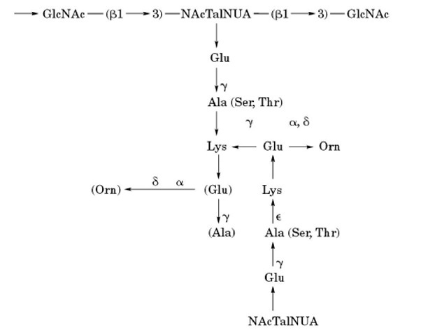
This type of cell wall is present in few Archaea, eg Methanobacterium formicicum, containing pseudomurein (1). The glycan strands of pseudomurein are composed of alternating b-N-acetyl-L-talosamine-uronic acid b-1 ^ 3 N-acetyl-D-glucosamine and the tetrapeptide contains L-amino acids (Fig. 2).
Figure 2. Pseudomurein in some methanogenic Archaea. GlcNAc, N-acetyl-D-glucosamine; NAcTalNUA, b-N-acetyl-L-talosamine uronic acid; Orn, ornithine; Ser, serine; Thr, threonine; Ala, alanine; Lys, lysine; Glu, glutamic acid.
![Schematic structure of polymers in cell walls of Gram-positive bacteria. A. Peptidoglycan with an polyglycine interpeptide bridge, [Gly]5 in Staphylococcus aureus. B. Teichoic acid of the ribitol-phosphate type. C. Lipoprotein with fatty acids (Rx to R3) linked directly or via glycerol to the terminal cysteinyl residue. Schematic structure of polymers in cell walls of Gram-positive bacteria. A. Peptidoglycan with an polyglycine interpeptide bridge, [Gly]5 in Staphylococcus aureus. B. Teichoic acid of the ribitol-phosphate type. C. Lipoprotein with fatty acids (Rx to R3) linked directly or via glycerol to the terminal cysteinyl residue.](http://what-when-how.com/wp-content/uploads/2011/05/tmp3639_thumb_thumb.jpg)

