Peptides, sometimes referred to as oligopeptides, are oligomers in which the repeating units are amino acids. Peptides have a defined sequence of amino acids that are linked together by formation of peptide bonds. In contrast to polypeptides and proteins, peptides consist of a small number of amino acids. The distinction between a peptide and a polypeptide is somewhat arbitrary, but generally a peptide has between 2 and 50 amino acid residues. A peptide of two amino acids may be more specifically referred to as a dipeptide, three repeating units as a tripeptide, four repeating units as a tetrapeptide, and so on.
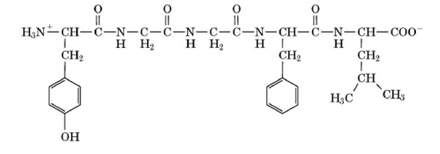
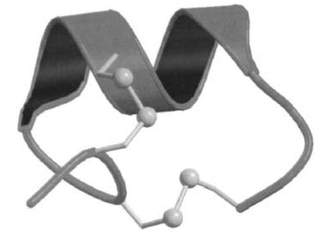
Most peptides are unstructured, described as having a random coil conformation, but others have highly ordered secondary and tertiary structure similar to that observed in larger proteins. The more structured peptides often incorporate disulfide bonds. Many peptides have important biological functions. For example, the pentapeptide leucine-enkephalin (Fig. 1) is a neurotransmitter with opiate-like activity; the hormone insulin is formed from two disulfide cross-linked peptide chains; and the major toxins from venomous spiders and cone shells are disulfide-rich peptides (Fig. 2). Naturally occurring peptides are usually produced by hydrolysis or cleavage of precursor proteins, through the action of peptidases or proteinases.
Figure 1. Chemical structure of the pentapeptide Leu-enkephalin, which has the sequence Tyr-Gly-Gly-Phe-Leu.
Figure 2. Schematic representation of the structure of the 16-residue a-conotoxin PnIA (1), showing the backbone and disulfides. The two turns of a-helix are depicted as a coil. The two disulfide bonds are shown in ball-and-stick representation. This figure was generated using Molscript (2) and Raster3d (3, 4).


