The past decade has witnessed enormous progress in understanding the molecular mechanisms that control the development of imaginal discs in insects. Lessons obtained from the studies of imaginal discs are being used to unravel mechanisms underlying vertebrate development, because many genes and developmental pathways are conserved throughout evolution.
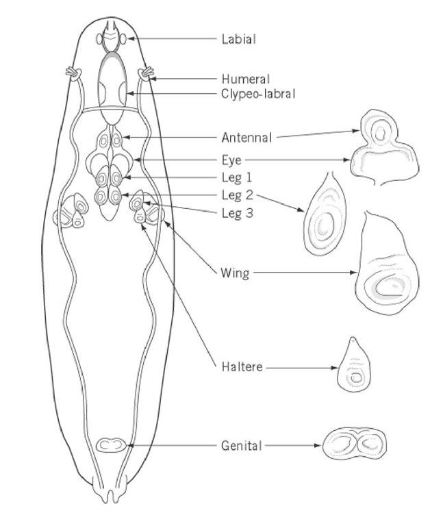
The development of imaginal discs is a feature unique to the holometabolous insects (beetles, moths, flies, and butterflies). The life cycles of holometabolous insects include four stages: embryo, larva, pupa, and adult (also known as imago). Development through these stages does not occur simply by increasing the sizes of exiting structures; instead, it involves a dramatic transformation from larva to adult. The transformation is achieved by the replacement of larval tissues with a special population of cells, known as imaginal discs, which form sac-like structures residing inside the larva. Imaginal discs have been studied most extensively in the fruit fly Drosophila melanogaster (1-4). There are 10 major pairs of imaginal discs, which give rise to adult cuticular structures of the head and the thorax, and a single genital disc, which forms the adult genitalia. The location of the imaginal discs in the mature larva (third instar stage) is illustrated in Figure 1. Studies in Drosophila have shown that imaginal discs are initially set aside during embryogenesis, apart from most embryonic cells that will contribute to the larva. In the embryo, the imaginal disc precursor cells can be visualized by the expression of molecular markers or by their particular sizes, shapes, and movement. The imaginal discs appear in the newly hatched larva as local thickenings of the epidermis and, in the case of the wing disc, contain around 40 cells (5). Subsequently, the imaginal disc cells proliferate rapidly during larval stages. By the end of the larval stage, the largest imaginal disc, that of the wing, contains approximately 50,000 cells (6). During pupation, the imaginal disc cells go through morphogenetic movements, in response to a pulse of the molting hormone ecdysone, to give rise to specific adult structures, accompanied by the breaking-down of the larval tissues.
Figure 1. Locations of imaginal discs in a third instar larva, and the morphology of several major imaginal discs. There are 10 major pairs of imaginal discs, which give rise to adult cuticular structures of the head and the thorax, and a single genital disc, which forms the adult genitalia.
The molecular studies of imaginal discs in Drosophila have been addressing the following major questions. First, how are the imaginal disc precursor cells set aside (or established) during embryogenesis? Second, how is the growth of imaginal discs in the larva regulated? Third, how do developmental patterns form in the mature imaginal disc along its anterior-posterior, dorsal-ventral, and proximal-distal axes? And fourth, how is the identity of a given imaginal disc specified? The following is a brief summary of our current understanding on these topics.
1. Establishment of the Imaginal Disc Precursor Cells in the Embryo
The fate of imaginal disc precursor cells is determined by intersecting anterior-posterior and dorsal-ventral signals in the embryo. These signals are provided by two sets of patterning genes along the embryonic anterior-posterior and dorsal-ventral axes. Along the anterior-posterior axis, a regulatory hierarchy involving gap genes, pair-rule genes, and segment polarity genes determines the different cell fates. In particular, the activities of several segment polarity genes, including wingless (wg), engrailed (en), patched (ptc), and hedgehog (hh), are required for the consolidation of the parasegment boundaries, which appear during early development of the embryo (7, 8). Among these genes, wg, encoding a secreted molecule of the Wnt family of proteins (9), is required for the establishment of the thoracic wing and leg discs, which originate from primordia spanning the parasegment boundaries (10, 11). Like the anterior-posterior axis, the embryonic dorsal-ventral axis is also patterned by a hierarchy of maternal and zygotic genes. In particular, the decapentaplegic (dpp) protein, a member of the transforming growth factor b (TGF-b) gene family of signaling molecules (12), and other molecules in the dpp signaling pathway function to specify dorsal cell fates (13, 14). In contrast, the DER protein, the Drosophila EGF receptor homologue, and other genes in the DER signaling pathway are required to specify ventral cell fates (15, 16). The positions of the thoracic disc precursor cells along the dorsal-ventral axis are determined by the combined repressing activities of the dpp and the DER proteins, which limit the thoracic disc precursor cells to lateral positions of the embryo (17). Therefore, the thoracic disc precursor cells are established by the intersecting signals of Wg along the anterior-posterior axis, and dpp and DER along the dorsal-ventral axis.
2. Regulation of the Imaginal Disc Growth in the Larva
As mentioned above, imaginal discs increase their number of cells by thousands of fold during larval development. Misregulation of disc growth can result in imaginal discs with abnormal sizes and morphology. Genetic studies have identified a large number of mutants that are either defective or excessive in disc growth. The disc overgrowth mutants are of particular interest, because they may provide genetic models for tumor suppressor genes in humans. Recessive mutations in several dozens of genes have been identified that cause two types of disc overgrowth phenotypes: hyperplasia and neoplasia (18-20). Hyperplastic discs retain the normal epithelial organization and the capacity to differentiate, whereas the neoplastic discs do not. These several dozens of genes are known as tumor suppressor genes in Drosophila, and over 10 of them have been characterized molecularly. For example, one of these genes, lethal(1)discs large (dlg), is the first identified member of a evolutionary conserved family of proteins, called membrane-associated guanylate kinase homologues (MAGUKs) (21). In Drosophila, dlg is localized at and required for the formation of septate junctions in epithelial cells, and loss of dlg function causes epithelia to lose their organization and overgrow (22). Further studies of these Drosophila tumor suppressor genes are likely to shed light on general mechanisms in the regulation of cell proliferation.
3. Pattern Formation in the Imaginal Discs
3.1. Anterior-Posterior Patterning
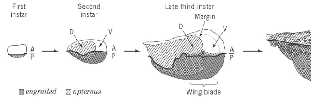
Previous studies have shown that imaginal discs are subdivided into distinct cell populations, called compartments (23, 24). The compartmental boundary serves as a line of lineage restriction, and cells in one compartment do not intermingle with cells in the other. The anterior and posterior compartments of the thoracic discs can be traced back to the embryonic stage when the disc precursor cells are specified initially along the anterior-posterior axis of the embryos (25). In these discs, the posterior compartment is defined by the expression of the transcription factor engrailed (en) (26), which activates the expression of hedgehog (hh) in the same compartment (27, 28). In the wing disc, the hedgehog protein synthesized in the posterior compartment diffuses a short distance into the anterior compartment to induce the expression of dpp in a stripe of cells just anterior to the anterior-posterior boundary (27, 29). The decapentaplegic protein in turn acts as a long-range morphogen to organize the growth and patterning of the whole wing (30, 31) (Figure 2). The leg disc is very similar to the wing disc, except that in the leg disc the induction of dpp by the hedgehog protein is limited to the adjacent dorsal anterior cells, whereas the adjacent ventral anterior cells are induced by the hedgehog protein to express wg (32, 53). In addition, dpp and wg mutually repress the transcription of each other (Figure 3) (34, 35). The unpaired genital disc consists of three primordia, the female genital, the male genital, and the anal primordia. Each primordium of the genital disc is divided into anterior and posterior compartments. Here, hh, dpp, and wg are used to pattern each primordium in a manner similar to how they function in the leg disc (36, 37). In contrast to the wing, leg, and genital discs, the eye disc is not divided into any lineage-restricted compartments. Pattern formation in the eye disc is accompanied by movement of the morphogenetic furrow (MF), a morphological distinguishable indentation, which sweeps across the eye disc from posterior to anterior. Cells anterior to the MF are undifferentiated, whereas cells posterior to the MF undergo cellular differentiation (37). The initiation of eye disc differentiation is regulated by interactions between the positive regulator dpp and the negative regulator wg. Subsequently, the progression of differentiation requires hh. Despite the lack of the anterior and posterior compartments, the regulatory relationships between hh, dpp, and wg are retained in the eye disc. It has been shown that dpp and wg suppress the expression of each other, and the hedgehog protein produced by cells posterior to the MF induces dpp expression within the MF (39, 40).
Figure 2. Axis formation and gene expression in the developing wing. In the first instar larva, the wing disc has already posterior compartment is identified by the expression of engrailed (26). In the second instar larva, the dorsal-ventral axi identified by the expression of apterous (33). The dorsal-specific expression of apterous is maintained during third larva begins to be expressed in a region slightly anterior to the anterior-posterior boundary (87). The dorsal and ventral surface of the late third instar disc: the perspective ventral surface folds under the dorsal one, while the wing blade unfolds and l wing blade region (X) is placed at the tip of the wing.
Figure 3. Spatial expression and regulatory interaction between hedgehog and other genes in the leg imaginal disc. The ] cells diffuses across the anterior-posterior compartment boundary; it induces the adjacent dorsal anterior cells to express anterior cells to express wingless (32, 55). The central region of the disc, where decapentaplegic and wingless are coexpi and aristaless and defines the distal tip of the leg (68-70).
Several other components of the hedgehog signal transduction pathway have been identified. Positively-acting components of the pathway include the seven transmembrane protein smoothened (41), the kinase fused (42), and the transcription factor cubitus interruptus (43); negatively-acting components of the pathway include the transmembrane protein patched (44, 45), protein kinase A (46-49), and the kinesin-related protein costal2 (50). The current model suggests that the patched and smoothened proteins constitute the receptor for the hedgehog signal (51). The cubitus interruptus protein, which is expressed only in the anterior cells due to the repression by the engrailed protein in the posterior compartment, forms a protein complex with the fused and costal2 proteins (50, 52). In some anterior cells, the cubitus interruptus protein is cleaved to generate a truncated form that translocates to the nucleus and represses dpp transcription. The hedgehog protein inhibits the proteolysis of the cubitus interruptus protein, leading to the expression of dpp in a stripe of anterior cells adjacent to the anterior-posterior boundary (53).
3.2. Dorsal-Ventral Patterning
During the second instar larval stage, the wing disc is also divided into dorsal and ventral compartments (23). The dorsal-ventral compartmental boundary lies at the future wing margin, which separates the upper surface of the wing from the lower one. Wing formation depends on the interactions between the dorsal and ventral cells (54). The dorsal selector gene apterous (ap), which encodes a LIM homeodomain protein, is expressed and required in the dorsal cells (33, 55-57), and it activates the expression offringe (fng) in these cells (58). The fringe protein acts through two ligands of the Notch receptor, Serrate and Delta (59-63), to restrict the expression of downstream genes wg and vestigal ( vg) along the dorsal-ventral boundary, which in turn coordinate wing growth and patterning (64-66).
3.3. Proximal-Distal Patterning
Besides the anterior-posterior and dorsal-ventral axes, appendages (wing and leg) also possess a proximal-distal axis. It appears that interactions between the anterior-posterior and dorsal-ventral axes of the disc are critical for patterning along the proximal-distal axis (67). In the leg, the hedgehog protein secreted from the posterior compartment induces the adjacent dorsal anterior cells to express dpp and ventral anterior cells to express wg. The region where wg and dpp are coexpressed expresses Distal-less and aristaless, which defines the most distal part of the leg (6871). Accordingly, a secondary proximal-distal axis can be induced when a new intersection point of wg and dpp expression is generated in the leg disc. Similarly in the wing disc, the intersection of dpp expression along the anterior-posterior boundary and wg-vg expression along the dorsal-ventral boundary corresponds to the distal tip of the wing.
4. Specification of the Imaginal Disc Identity
Although patterning of all imaginal discs share some common features, the identity of an individual imaginal disc is specified by the homeotic and other selector genes. Homeotic genes in the Bithorax and Antennapedia complexes are involved in the specification of head and thoracic discs (72). For example, Ultrabithorax (Ubx) is expressed in the haltere, but not in the wing disc, and specifies the haltere fate (73-75). Loss of Ubx function in the haltere results in a haltere-to-wing transformation, generating the famous four-winged fly (76). The homeotic gene Antennapedia (Antp) specifies the leg disc, and ectopic expression of Antp in the eye-antenna disc results in antenna-to-leg transformation (77, 78). However, homeotic genes are not the only genes that are used to specify disc identity. The Drosophila Pax family gene eyeless (ey), which encodes a protein containing a paired domain and a homeodomain, is required for the determination of the eye disc fate (79). Ectopic expression of ey can induce ectopic eye development in other imaginal discs (80). Several genes have been shown to act downstream of ey in this process, including eyes absent, sine oculis, and dachshund (81, 82).
Studies of imaginal disc development in Drosophila have shed new light on various aspects of vertebrate development. For example, gene expression and functional studies suggest a striking similarity between fly appendages and vertebrate limbs (32, 67, 83, 84). The dorsal-ventral boundary of the wing disc is analogous to the apical ectoderm ridge (AER), which is a major signaling center for the developing vertebrate limb. The posterior compartment of the wing disc is analogous to the zone of polarizing activity (ZPA), the source of the morphogen, sonic hedgehog, that patterns the anterior-posterior axis of the vertebrate limb bud. In addition, several key regulatory genes in patterning imaginal discs have been implicated in human diseases. For example, mutations in the human patched gene are responsible for basal cell carcinoma, a common form of human cancer (85).


![Spatial expression and regulatory interaction between hedgehog and other genes in the leg imaginal disc. The ] cells diffuses across the anterior-posterior compartment boundary; it induces the adjacent dorsal anterior cells to express anterior cells to express wingless (32, 55). The central region of the disc, where decapentaplegic and wingless are coexpi and aristaless and defines the distal tip of the leg (68-70). Spatial expression and regulatory interaction between hedgehog and other genes in the leg imaginal disc. The ] cells diffuses across the anterior-posterior compartment boundary; it induces the adjacent dorsal anterior cells to express anterior cells to express wingless (32, 55). The central region of the disc, where decapentaplegic and wingless are coexpi and aristaless and defines the distal tip of the leg (68-70).](http://what-when-how.com/wp-content/uploads/2011/05/tmpFC171_thumb_thumb.jpg)
