Biomedical Engineering Reference
In-Depth Information
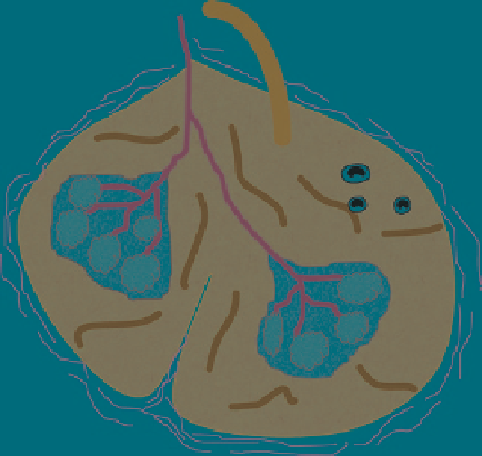
Vein
Artery
Monocytes
Vascular sinusoid
Red pulp
Lymphoid follicule
White pulp
Capsule
Fig. 4
Representation of the basic structure of the spleen containing red and white pulp
This function takes place in the red pulp of the spleen that comprises the majority
of the organ's structural surface area (Fig.
4
). The spleen is the largest organ
within the lymphatic network and functions to filter blood in a similar manner
to the way lymph nodes filter lymph. Blood enters the spleen via the splenic
artery that extends into the white pulp and branches into the venous sinuses that
remove damaged red cells. Splenic cords extend between venous sinuses that
contain reticular connective tissue, macrophages and lymphocytes that act to
remove dead cells and foreign material from the blood. The spleen also acts as
a reservoir for red cells that can be added to the blood via splenic contractions
when needed.
The spleen also has clusters of white pulp that are responsible for producing,
storing and exchanging lymphocytes with the blood. Therefore, nanomedicines
circulating in the blood can be removed either via macrophages and lymphocytes
in both the red and white pulp of the spleen. As an example, the biodistribution of
polylysine dendrimers conjugated with both polyethylene glycol and methotrexate
display more avid uptake into the spleen than in the liver of athymic nu/nu rats that
have a reduced number of T cells (Kaminskas et al.
2009a
). Macrophages in the
spleen likely act in this instance to compensate for the loss of T cell activity by
becoming more phagocytically active. Also, the relative biodistribution of lipo-
somes into liver and spleen varies according to composition, where liposomes
composed of egg phosphatidylcholine show roughly equivalent uptake into liver
and spleen, whereas liposomes containing phosphatidylserine or dipalmitoylphos-
phatidylglycerol (DPPC) are more avidly taken up into the spleen (Oussoren and
Storm
1997
).


Search WWH ::

Custom Search