Biomedical Engineering Reference
In-Depth Information
Branch of
Portal Vein
Bile duct
Bile duct
Hepatocytes
Stellate cell
Sinusoid
Kupffer cell
Sinusoidal
Endothelial cell
Sinusoid
Branch of
Hepatic Artery
Hepatocytes
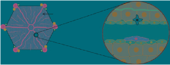
Fig. 3
Diagram of the basic lobular structure of the liver (
left
) and of the microstructure surrounding
the sinusoids (
right
)
The major microstructural unit of the liver is the lobule (Fig.
3
), a hexagonal
structure that contains a central vein with blood tracts that protrude from the main
structure (called the sinusoids that are lined with a thin layer of sinusoidal endothe-
lial cells) and branches of the portal vein, hepatic artery and bile ducts at each corner
of the hexagonal lobule. The major cell type that resides between the sinusoids are
the hepatocytes. These are the major metabolic cells of the liver and are storage sites
for a number of essential nutrients that occur in excess in the blood, such as iron and
glycogen. The hepatocytes occur in a single layer between the liver sinusoids and
bile cannaliculi and are responsible for absorbing drugs and materials from the blood
and processing them for secretion into the bile ducts. Thus, the excretion of nano-
medicines via the bile and subsequently via the feces requires initial uptake via the
hepatocytes. Hepatic stellate cells also lie in between clusters of hepatocytes. These
cells are the fat storage cells of the liver. Finally, Kupffer cells (the major phagocytic
cells of the liver that account for approximately 2% of the liver volume) lie within
the sinusoids. Their residence at this site optimizes their exposure to blood patho-
gens, foreign material and more importantly to this chapter, nanomedicines. Blood
entering the liver from the hepatic artery and the portal vein flow towards the lobule
via their branches, through the sinusoids where the blood is 'cleaned' and into the
central vein that pools detoxified or cleaned blood into the hepatic vein. There are
also two distinct types of Kupffer cells in the liver. The smaller, more immature cells
are mainly located in the centrilobular region. The larger, more mature cells that play
a more prominent role in phagocytosis, and hence are of more importance in the
uptake of particles from the blood by the liver, are located in the periportal region.
2.2
The Spleen
The spleen is important in generating blood cells during fetal development, but it
is not a vital organ in adults. It does, however, function in addition to the liver to
destroy and remove damaged or aged red blood cells from the systemic circulation.


Search WWH ::

Custom Search