Biomedical Engineering Reference
In-Depth Information
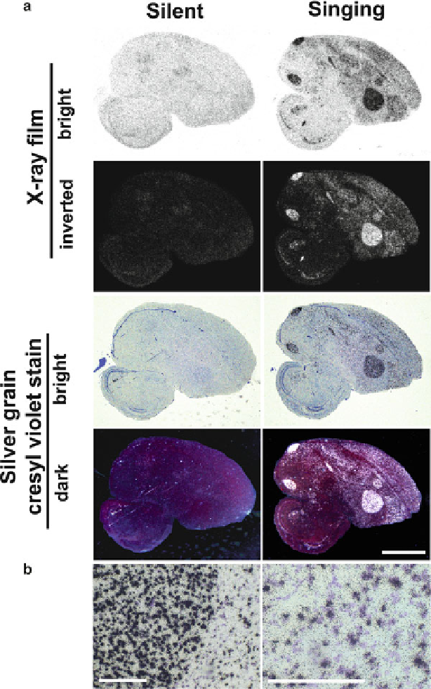
Fig. 9.7
Photo images of radioactive signals. (
a
) whole brain in-situ hybridization images under
silent and singing conditions, using X-ray fi lm images under bright light (top panels; black = mRNA
signal), inverted X-ray fi lm images (2nd low; white = mRNA signals), silver grain dipped-cresyl
violet stained images under regular bright light (3rd low; black = mRNA signals, blue/purple =
cresyl violet stained cells), and silver grain dipped-cresyl violet stained images under polarized light
in dark fi eld (4th low; white = mRNA signals, purple/red = cresyl violet stained cells). Scale bar =
2mm. (
b
) Higher magnifi cation of silver grain dipped-cresyl violet stained images under regular
bright light. Developed silver grains are observed as black dots. Scale bars = 200µm