Information Technology Reference
In-Depth Information
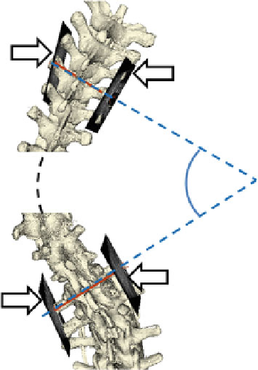
Fig. 18 Spinal curvature
measurement using four
landmark points from four
tracked ultrasound snapshots
(marked by white arrows).
The 3D spine model
illustrates the measurement
principle, but it is not
available in the clinical setting
used for ultrasound-based measurement that is used for X-ray measurement. The
vertebral end-plates cannot be seen in ultrasound due to the acoustic shadow of the
lamina and vertebral processes. Anatomical features that are accessible by ultra-
sound imaging and also visible in X-ray are transverse processes (Fig.
19
).
Two potential advantages of using tracked ultrasound for spine curvature angle
measurements are safety and accessibility. Radiation-free monitoring method in
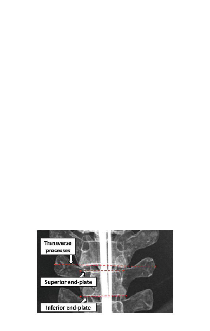
Fig. 19 Anatomical features for spinal curvature measurement. Superior and inferior end-plates
are conventionally used in radiographic measurements. The transverse processes are also visible in
ultrasound