Information Technology Reference
In-Depth Information
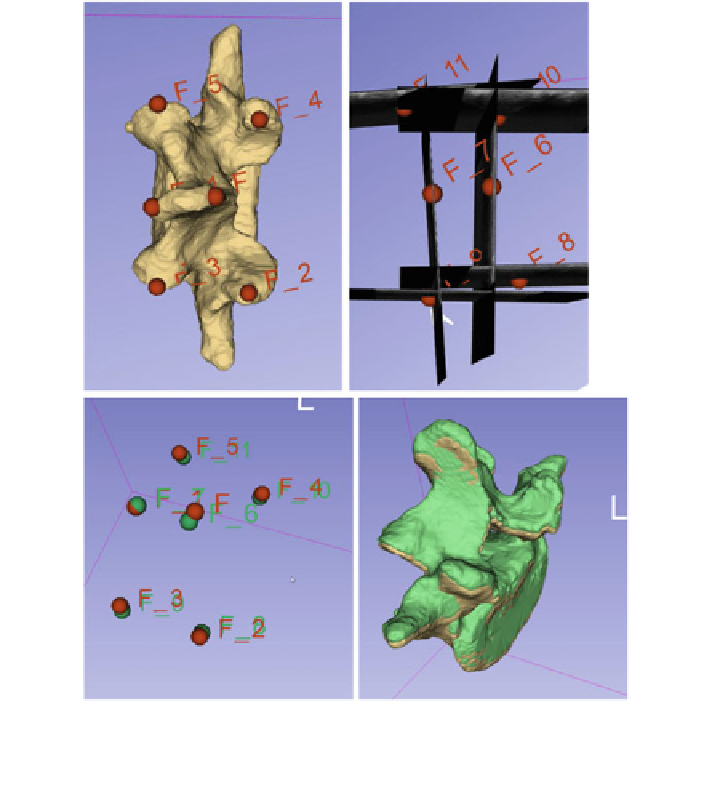
Fig. 17 Landmarks de
ned for registration on the CT-derived model of a lumbar vertebra (top left
image), and the same landmarks de
ned on tracked ultrasound snapshots (top right image). The
two sets of landmarks are registered (lower left image), and the registered vertebra position (green)
is localized close to the ground truth position (yellow) in the lower right image
Tracked ultrasound offers accurate spatial localization of vertebra landmarks
visible on ultrasound images. These landmarks are suitable for measurement of
spinal curvature and vertebra rotation without ionizing radiation. Spinal curvatures
are measured between two vertebrae that are rotated in the coronal plane at the
largest angle. The angle is de
ned between two lines in the coronal plane. Both
lines can be de
ned by two symmetric points on each vertebra. The points can be
transverse processes on tracked ultrasound snapshots, as these points are visible on
ultrasound images along the entire spinal column (Fig.
18
).
Tracked ultrasound technique can provide as accurate spinal curvature mea-
surements as X-ray images [
26
]. Although this method needs further clinical test-
ing, as the conventional anatomical landmarks, the vertebral end-plates, cannot be