Biomedical Engineering Reference
In-Depth Information
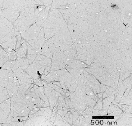
Figure8.7
TEMmicrographofchitinnanocrystalsfromshrimpshellsformedafterhydrolysis
andmechanical dispersion. Magnification shown is (
10,000). Reprintedwith permission
from(23).Copyright(2007),AmericanChemicalSociety.
×
structural domains within the microfiber. Peaks arising due to C2, C3 and C5 atoms
occur between 72 and 79 ppm and finally the C6 peak has a chemical shift value of
65 ppm. The absence of any aromatic signals between 110 and 140 ppm clearly indicated
that the lignin component present in bagasse had been successfully eliminated as a result
of the alkali treatment and the subsequent bleaching process.
The earliest published work on the solid-state NMR spectra of cellulose showed two
peaks in the chemical shift range 80-92 ppm and these have been assigned to the C4
carbon atom (24, 25). A relatively sharp peak was assigned to crystalline regions, and
a relatively broad peak was attributed to the crystallite surfaces and the amorphous/
disordered domains.
Previous assignments by Newman (26, 27), Horii (28), and Iversen (29) have corre-
lated the solid-state NMR spectra of cellulose with its structure and morphology. A weak
shoulder on the C6 peak, between 63 and 65 ppm, has been attributed to the amorphous
and disordered component in cellulose. This includes the surface of crystal domains
since they need not participate in the symmetry of the crystal. The
13
C NMR spectra of
the cellulose fibers before and after hydrolysis have some very distinct differences:
1. The signal assigned to the C4 peak changed dramatically. There was a significant
difference between the peak profiles of the crystalline and the amorphous compo-
nents attributed to signals in this region, (80-92 ppm) before and after hydrolysis and
mechanical shearing. The unhydrolysed cellulose fibers exhibited roughly equal con-
tributions from the crystalline and the amorphous domains (Figure 8.9a). Newman
and his co-workers have used curve-fitting specific assignments to the signals arising
from crystallite interiors, crystallite surfaces as well as the amorphous regions. No
such attempt was made in our case because of the large number of scans required
Search WWH ::

Custom Search