Chemistry Reference
In-Depth Information
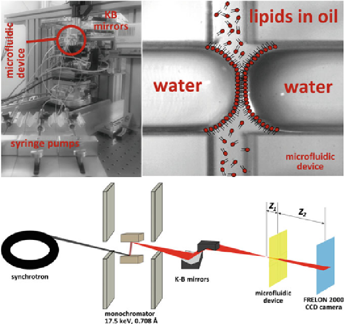
Fig. 4.1
Aschematic of the synchrotron setup, at the ID22 (ESRF) beamline, for imaging amicroflu-
idic lipid bilayer. Amicrofluidic device in which a lipid bilayer can be formed by two water droplets
suitably covered by a monolayer of lipid
the formation of convex water interfaces (also called “water fingers”). Subsequently,
the oil flow is stopped and the water fingers are slowly brought into contact in
the channel cross region. When the two monolayers approach each other, intense
Fresnel fringes appear in the visible light microscope before the monolayers fuse
and form a bilayer. The size of the bilayer patch can be controlled by variation
of the pressure in the water channels. Presence of a single bilayer is checked by
insertion of electrodes in the aqueous channels to measure the capacitance of the
interface. The orientation and geometry of the microfluidic lipid bilayers shows a
great advantage compared to bulged black lipid membranes that were investigated in
[
10
,
11
]. As the phase contrast signal of a transparent object increases proportionally
to the path length
L
of the X-rays inside the sample, the microfluidic bilayers offer an
improved geometry for imaging experiments.
L
is in the range of a few micrometers
in the case of a thin, bulged BLM. The diameter of a bilayer can equal the depth
of the channel when it spans it fully. Consequently, the penetration depth becomes