Chemistry Reference
In-Depth Information
changes the membrane distance from 5-28nm to below that required for SNARE
complex formation (8nm at normal ionic strength [
36
]) such that membrane fusion
can occur.
3.4 Summary and Outlook
In this chapter we investigated SNARE mediated membrane fusion under conditions
where electrostatic interactions between the component membranes are not screened.
In order to study these interactions, we developed a microfluidic technique based on
a standard FRET assay for membrane fusion. Our data show that in a simple recon-
stituted system, key elements of synaptic transmission can be mimicked if SNARE
nucleation is prevented by keeping themembranes apart before
Ca
2
+
-triggering. The
charged lipids that constituting the bilayers result in repulsive interactions that keep
the membranes too far for even SNARE nucleation to occur. However, even under
such conditions, synaptotagmin-1, a
Ca
2
+
trigger for membrane fusion, tethers the
two lipid membranes, although the SNARE proteins are fully active. Upon addi-
tion of
Ca
2
+
, the binding of
Ca
2
+
to synaptotagmin-1 results in a conformational
change of the protein that brings lipid bilayers close enough for SNARE nucleation
to occur and thus for membrane fusion to proceed (Fig.
3.14
). While we were able to
reproduce, in vitro, the massive increase of membrane fusion triggered by
Ca
2
+
,the
condition of low ionic strength of the buffer is not a relevant physiological condition.
However, it is conceivable that proteins residing in the space between the vesicle
and the plasma membrane can fulfill the condition of keeping the membranes too far
for fusion to readily occur. Considering that both synaptic vesicles and release sites
are indeed crowded with membrane proteins [
41
], such a scenario is not unlikely.
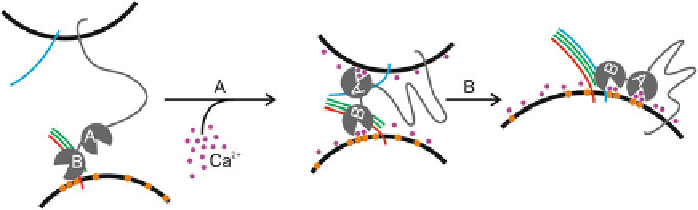
Fig. 3.14
Model for synaptotagmin-1 mediated lipid mixing. In the absence of
Ca
2
+
,
synaptotagmin-1 (
grey
) tethers to anionic membranes, and particularly to Pi(4,5)
P
2
(
orange
)via
its polybasic lysine patch. The distance is too far for SNARE formation to occur (synaptobrevin-2:
blue
; Syntaxin-1A:
red
; SNAP-25:
green
). (Step A) In the presence of
Ca
2
+
(
purple
), a transitional
conformation change occurs and synaptotagmin-1 binds the membrane via its calcium binding
pockets and basic residues on the C2AB-domain. This drives the membranes together and SNARE
complex formation can occur (Step B) The SNARE complex formation drives membrane fusion