Biomedical Engineering Reference
In-Depth Information
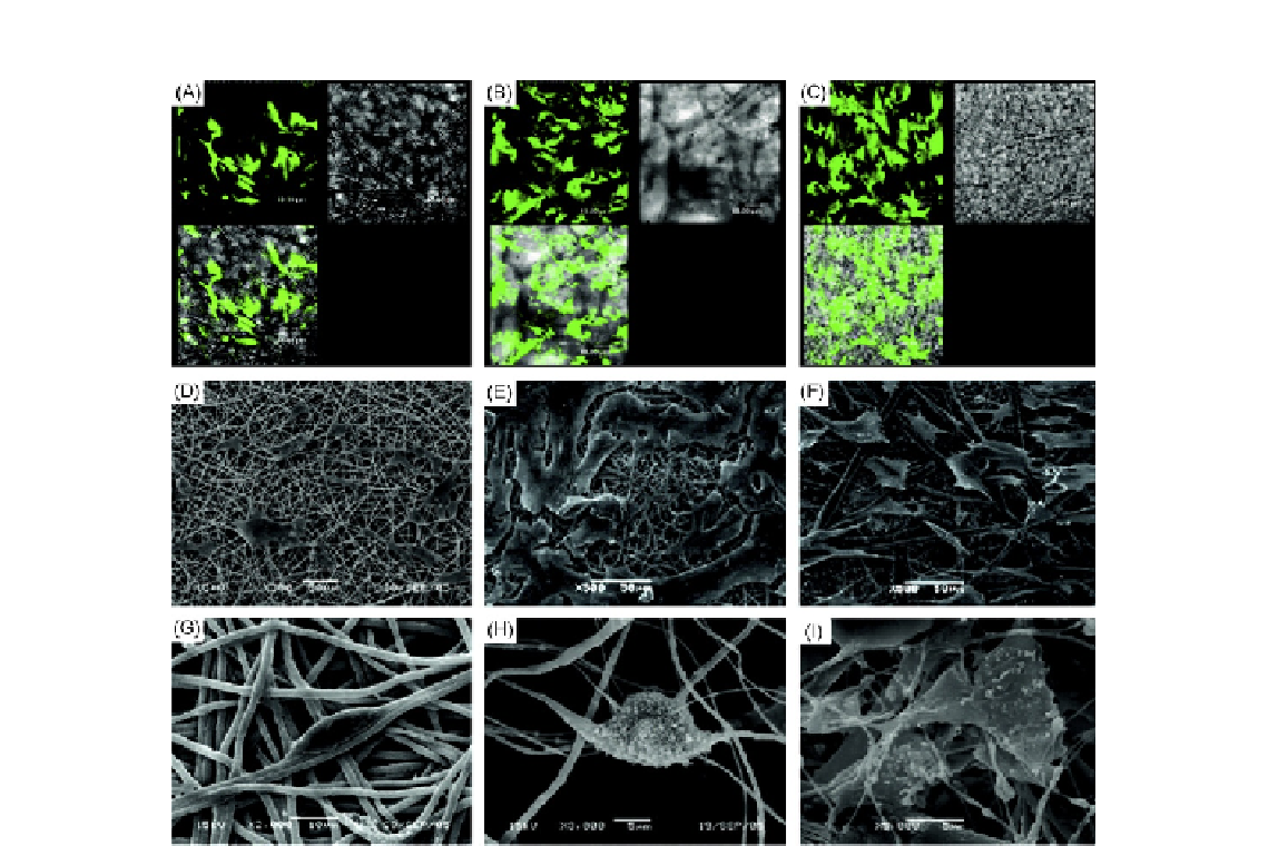
FIGURE 10.6
Morphology observation of PDLCs cultured on three kinds of membranes: (A-C) represent confocal laser
microscope images of PDLCs on PLLA, PLLA/HA, and PLLA/MWCNTs/HA membranes, respectively;
(D-F) represent scanning electron microscope (SEM) images of PDLCs on PLLA, PLLA/HA, and
PLLA/MWCNTs/HA membranes, respectively, at low magnification, and (G-I) represent them at high
magnification.
formed with a round or irregular shape (areas pointed by white arrow) and were stained into
homogeneous pink by hematoxylin/eosin, and osteoblast-like cells were well-arranged around
the bone-like tissues. Calcium deposits were confirmed in newformed bone-like tissue by aliza-
rin red staining. It was notable that abundant blood vessels were grown into the newformed tissues.
Osteocalcin, which was stained in brown, was detected in the cytoplasm and outside the cells (pointed
by white arrow). The results indicated that PLLA/MWCNTs/HA membranes were of good bio-
compatibility
in vivo
during the 4-week period, and human PDLCs seemed to function well on the
membrane.