Biomedical Engineering Reference
In-Depth Information
(A)
(B)
PLLA
PLLA/HA
PLLA/MWNTa/HA
TCPs
PLLA
PLLA/HA
PLLA/MWNTa/HA
TCPs
1
0.7
0.6
0.5
0.4
0.3
0.2
0.1
0
0.9
0.8
0.7
0.6
0.5
0.4
0.3
0.2
0.1
0
1
3
5
7
1
3
5
7
Day
Day
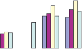
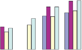
FIGURE 10.7
Effect of three kinds of membranes on the adhesion and proliferation of PDLCs (A) and GECs (B)
determined by MTT assay. Standard deviations are shown as bars.
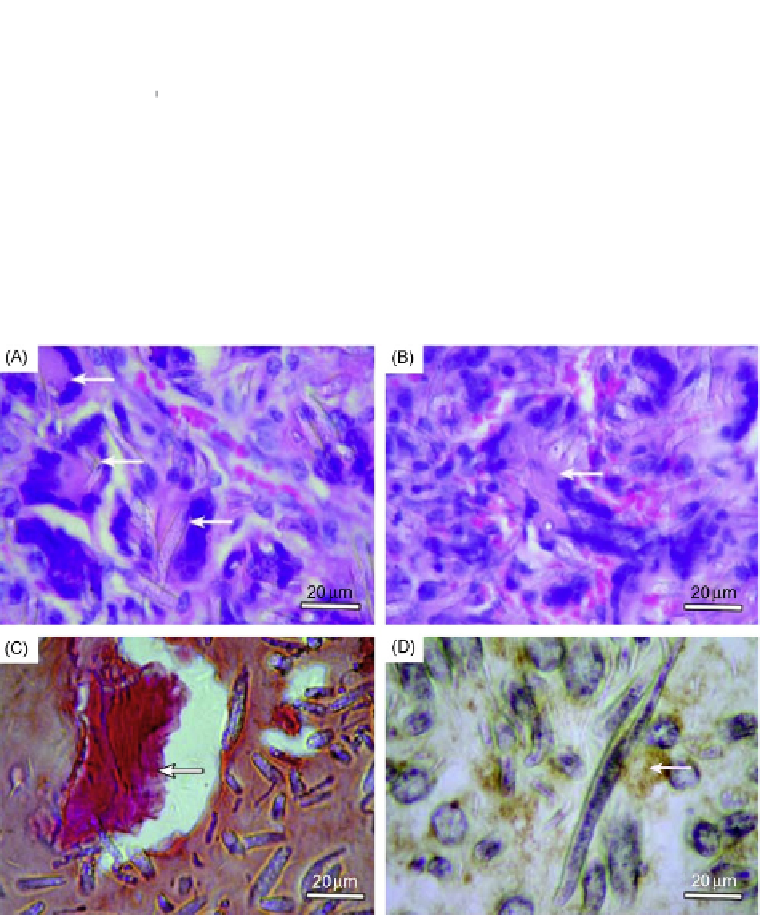
FIGURE 10.8
Histological examination of cell/membrane composites implanted into immunodeficient mice:
(A-C) show newformed bone-like tissues in round or irregular shape (white arrow), and
osteoblast-like cells were well arranged around bone-like tissues. Abundant blood vessels were
found in the implanted area. In (C), alizarin red staining confirmed calcium deposits (white
arrow) in new formed bone-like tissues. In (D), osteocalcin, which was stained in brown (white
arrow), was detected in the cytoplasms and outside the cells.