Biomedical Engineering Reference
In-Depth Information
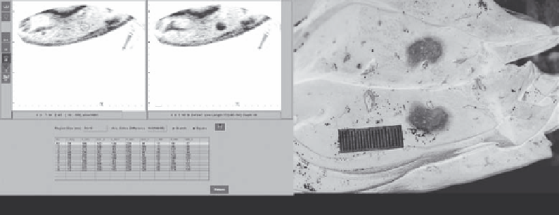
(a)
(b)
FIGURE 10.10
(a) Comparison of grayscale value of targeted liver tissue before (left) and after (right) HIFU exposure; (b) the range of hyper-
echoic area is good correspondence to the maximal area of coagulation necrosis.
The ultrasonic monitoring system is mainly used for locating
a targeted tumor, monitoring treated-tissue response, and con-
trolling the deposition of ultrasound energy during the HIFU
procedure. The treatment control system is composed of a treat-
ment planning system (TPS), digital evaluation of the grayscale
changes within the targeted tissues before and after HIFU expo-
sures, treatment data storage, and data analysis.
he
auxiliary system
includes a water management device
for degassing and temperature controlling and a safe protection
device. The water management device provides degassed water
as a coupling medium allowing ultrasonic propagation from
the transducer to the skin. Degassing is used to avoid skin burn
that is caused by air bubbles from the water gathered on the skin
surface (bubbles stop/scatter sound waves). The oxygen content
is kept at ≤3 ppm. As sound attenuation is very low in water,
the loss of ultrasound energy is not significant while the sound
waves travel through the water tank. The safe protection devices
ensure equipment stability and normal operation.
In addition, a telemedicine unit is embedded in the Haifu
system in order to let users share their treatment database with
other users or remote experts. It can also provide real-time con-
sultation and diagnostic services for cancer patients, and timely
repair and maintenance remotely.
system include superior image quality and anatomic detail and
real-time temperature measurement.
For US-guided HIFU systems, the evaluation of therapeutic
response is based on echo changes in the targeted tissues before
and immediately after HIFU exposures. Compared to US imag-
ing before HIFU, there is a strong echo from the targeted tissue
immediately after a HIFU exposure. With the extension of obser-
vation time, the echo gradually weakens, and the range of the
strong echo significantly decreases. Using proprietary software,
the strength of the echo is automatically analyzed in the targeted
area before and after HIFU exposures to derive the changes in
grayscale values. Coagulation necrosis is predicted based on
these changes. There is good agreement between the range
of the hyperechoic area and the maximal area of coagulation
necrosis (Figure 10.10). During the HIFU procedure, the gray-
scale changes are helpful in determining whether coagulation
necrosis occurs in the treated tissue. If the change cannot reach
a certain value after one HIFU exposure in terms of grayscale
differences, HIFU exposures are repeated until the grayscale
values reach the level of coagulation necrosis. Although it is still
unclear which mechanisms are involved in the development
of the hyperechoic area induced by HIFU exposures, acoustic
cavitation and tissue dehydration/boiling (evaporation) may be
major reasons responsible for hyperechogenicity.
10.3.2 US Imaging Guidance
As a noninvasive thermal ablation tool for tumor therapy, HIFU
is performed under image guidance. Either diagnostic ultra-
sound or MRI is clinically used to guide the HIFU treatment
procedure (depending on model). Both imaging modalities have
similar functions for real-time guidance, such as positioning
a target area, monitoring either temperature rise or grayscale
changes in the target, evaluating therapeutic response, and con-
trolling energy delivery. Benefits of a US-guided HIFU system
include low cost, real-time imaging, and sound channel vis-
ibility. In addition, there are no image artifact problems when
imaging moving internal organs (e.g., liver, kidney, and pan-
creas) due to respiration. Advantages of an MRI-guided HIFU
10.3.3 Quality assurance
The quality assurance of HIFU systems is very important to
ensure the safety of patients and the effectiveness of the treat-
ments. China has been a leader in developing and establishing
quality standards for clinical HIFU products. In September
2005, the national standard GB/T 19890-2005 “The measure-
ment of acoustic power and field characteristics in high inten-
sity focused ultrasound (HIFU)” was published by the Chinese
government, making it the first of its kind in the world. In
fact, it was published as a technical report by the International
Electrotechnical Commission (IEC/TR 62649) in April 2010.
China's SFDA issued the industry standards for HIFU therapy