Biomedical Engineering Reference
In-Depth Information
minimally invasive therapeutic technique for future cancer ther-
apy.
Ultrasonics Sonochemistry
, one of the international journals
in ultrasound, reported on the findings at this symposium that
patients with malignant tumors who were not suitable for sur-
gery and were treated by ultrasonic noninvasive methods have
survived for ten years. China is the only country in the world
that has provided such data owing to its long-term research and
clinical applications in this field
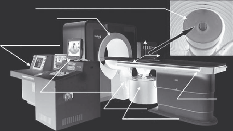
10.3.1 Main Components
The Ultrasound-Guided Extracorporeal HIFU system consists
of three main parts: the treatment platform, the central operator
console, and the auxiliary system (Figure 10.9). Each functional
module is described next.
he
treatment platform
includes a power source, an integrated
transducer, and a six-dimensional motion device for transducer
movement. The power source provides high-frequency electrical
signals that drive the HIFU transducer. The integrated trans-
ducer includes a 1.0 MHz HIFU transducer and a 3.5-5.0 MHz
B-mode ultrasound imaging probe, which is embedded in the
center of the HIFU transducer for real-time monitoring of the
HIFU procedure, including localization of the target, treatment
planning, observation of targeted tissue response in terms of tis-
sue grayscale changes, and control of ultrasound energy deliv-
ery. The integrated transducer is inside a rubber water tank filled
with circulating degassed and temperature-controlled water. The
transducer can move in six directions, including translational
motion in the x, y, and z axes, rotation of the imaging probe for
3D ultrasound imaging of the targeted area, and rotation of the
integrated treatment transducer along the x and y axes.
he
central operator console
includes an ultrasonic (imag-
ing) monitoring system and a treatment control system.
FIGURE 10.8
Model JM 2.5C HIFU system (Chongqing Haifu).
In 2005, Chongqing Haifu started an R&D project in collabo-
ration with Siemens Medical Systems to develop an MRI-guided
extracorporeal HIFU System (JM 2.5C HIFU System) shown in
Figure 10.8. The JM 2.5C system was fully integrated with a 1.5 T
MRI system (Symphony, Siemens, Germany). With the success-
ful development of the device, clinical trials in China have
recently been completed for the treatment of uterine fibroids.
The results show that the JM 2.5C system is safe, reliable, and
effective for the treatment of patients with large uterine fibroids.
Using the JM 2.5C system, a higher ratio of complete ablation
and a relief of symptoms related to fibroids have been observed in
patients during a 2-year follow-up, with a significant shrinkage
of the treated fibroids. These findings were reported at the First
International Clinical Symposium of Therapeutic Ultrasound in
October 2009. More than 120 clinicians and scientists from 19
countries attended this symposium, shared their clinical experi-
ences in HIFU therapy, and discussed the potential of HIFU as a
Integrated treatment transducer (diagnostic US probe
and HIFU transducer) in degassed water reservoir
γ-Motion device
Z
Treatment control system
X
Y
Ultrasonic monitoring system
Treatment bed
ψ-Motion device
3+1 D motion device
FIGURE 10.9
Haifu model JC focused ultrasound tumor therapy system.