Biomedical Engineering Reference
In-Depth Information
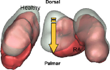
Fig. 5.4 Superior view of
the proximal row carpal
bones. The figure shows its
dislocation towards palmar
and ulnar directions. The RA
bones were in red and
transparent bones represent
the normal healthy bones
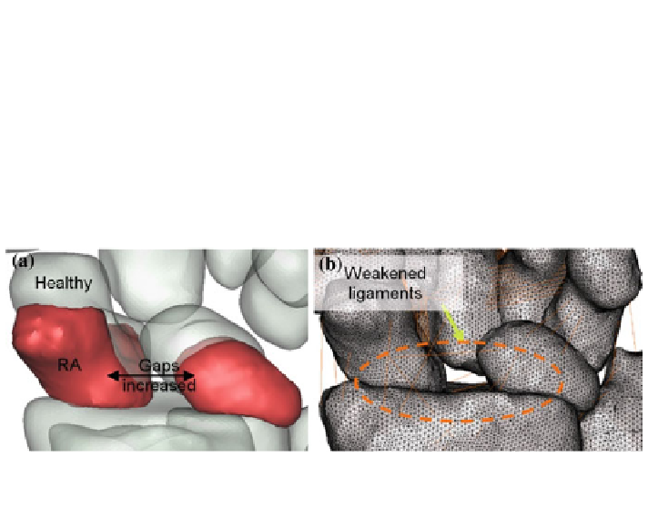
Fig. 5.5 The simulated SLD and SLAC were shown in (a) where the RA bones were in red and
the transparent bones represent the healthy bones. The simulated effect of weakened and torn
ligaments was also shown (b)
5.1.5 Simulation of Scapholunate Dissociation and Scapholunate
Advanced Collapse
The SLD was simulated by increasing distance between the scaphoid and the lunate,
from 1.98 to 6.51 mm. The worn and torn intrinsic ligaments as results from the
synovitis effect [
2
,
5
] were simulated by utilising one link. As stated by Trieb et al.
[
2
], the scapholunar and lunotriquetral ligaments are commonly effected as the
disease progresses, thus this condition subsequently destruct the scapholunate
articulation [
9
,
17
,
18
]. This circumstance was even worse as the high mobility of
the scaphoid has resulted in imbalance load transfer from the distal to the proximal
through the joint [
16
], and even pronounced as the capitate dissociates the scaphoid
and the lunate further. SLAC was then diagnosed as the disease progressed. This
condition incorporates the triquetrum, and its distance from the lunate was also
reduced. Again, the synovitis leads to the dislocation of the triquetrum and the lunate
towards distal ulnar. The simulated characteristic is as shown in Fig.
5.5
.
Search WWH ::

Custom Search