Biomedical Engineering Reference
In-Depth Information
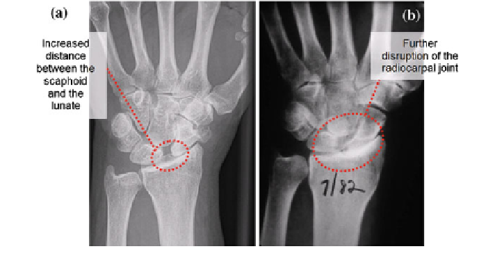
Fig. 3.3
Radiograph of the wrist with SLD (a)[
20
] and SLAC (b)[
7
]
to synovial expansion, results in ulnar translation and carpal supination. The
pathological process begins with inflammation of the synovial, affects commonly
at the ulnar side of the wrist joint [
10
,
12
]. It then spreads to adjacent area of the
wrist including the radiocarpal joint. The neighbouring tissues which consist of the
cartilages, ligaments and tendons degenerate subsequently. In severe cases, tendon
rupture occurs with a consequence of kinematic changes of the joint, resulting in
disruption of the periarticular bones and the articular surfaces [
9
,
19
]. On the
whole, these three symptoms have critically caused degeneration of both soft
tissues and bones, hence eventually mutilated and unstable wrist joint.
Trieb et al. [
13
] have successfully identified the pathophysiological character-
istics of the wrist with RA. Ligamentous laxity for both the intrinsic and extrinsic
ligaments has resulted in unphysiological bones movements. This was evident as
loss of tension of the radiotriquetral ligament caused dislocation of the carpus in
the ulnar direction. Scapholunar dissociation (SLD) due to the increased distance
between the scaphoid and lunate is primarily occur because of deteriorated
intrinsic ligaments: scapholunar and luno-triquetral which caused by synovial
inflammation [
20
] (Fig.
3.3
a). Progression of this SLD will also lead to a more
severe deformation of the joint, known as scapholunate advanced collapse (SLAC)
(Fig.
3.3
b).
Another pathophysiology characteristic is the dislocation of the proximal carpal
row in the ulnar and palmar directions. Ulnar dislocation of the bones was
attributed to the weakened radiotriquetral ligament and destructed capitolunate
joint, thus resulted in relatively greater load being transferred to the lunate. The
reduction of contact between the lunate and the radius was also found in rheumatic
wrist due to the dislocation of lunate in the ulnar direction. The scaphoid as the
most problematic carpal bone was commonly found to be dislocated palmarly due
to deteriorated radioscapholunate ligament. The impaction or loss of carpal height
was due to bone erosion and the unphysiological bones dislocations which worsen
Search WWH ::

Custom Search