Image Processing Reference
In-Depth Information
3
2
1
(a)
(b)
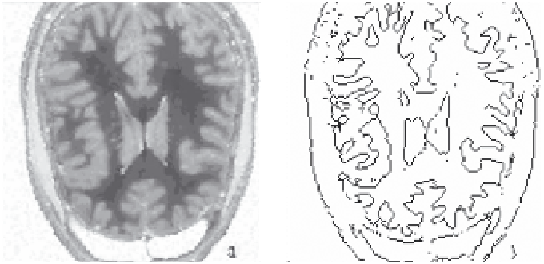
FIGURE 8.2
(a) Brain image and (b) edge-detected image. (Modified from Chaira, T. and Ray, A.K.,
Appl.
Soft Comput.
, 8(2), 919, 2007.)
8.3.2 Edge Detection Using the Median Filter
In this algorithm, the median filter is used to detect the edges of medical
images [6]. As medical images are not properly illuminated, direct edge
detection techniques sometimes do not work. So, to highlight the edges of
the image, edges are enhanced. In blood cell/vessel or nuclei images, nuclei/
cells/vessels are not clearly visible, making this technique beneficial. After
enhancing the image, edge detection techniques are applied.
An image (say
A
) of size
M
×
N
is initially fuzzified using the following
formula:
gg
−
(8.3)
min
μ
F
()
g
=
g
−
g
max in
where
g
is the grey level that ranges from 0 to
L
− 1
g
min
and
g
max
are the minimum and maximum values of the grey levels of
the image, respectively
Based on the fuzzy set, the membership degree of the intuitionistic fuzzy
image is calculated as [20]
(8.4)
λ μλ
λ
μ
(;)
g
=−−
11
(
(;))
g
IFS
F
and the non-membership degree is
λλ
(
+
1
)
ν
(;)( (;))
g
λ μλ λ
=−
1
g
,
≥
0
IFS
F
Search WWH ::

Custom Search