Image Processing Reference
In-Depth Information
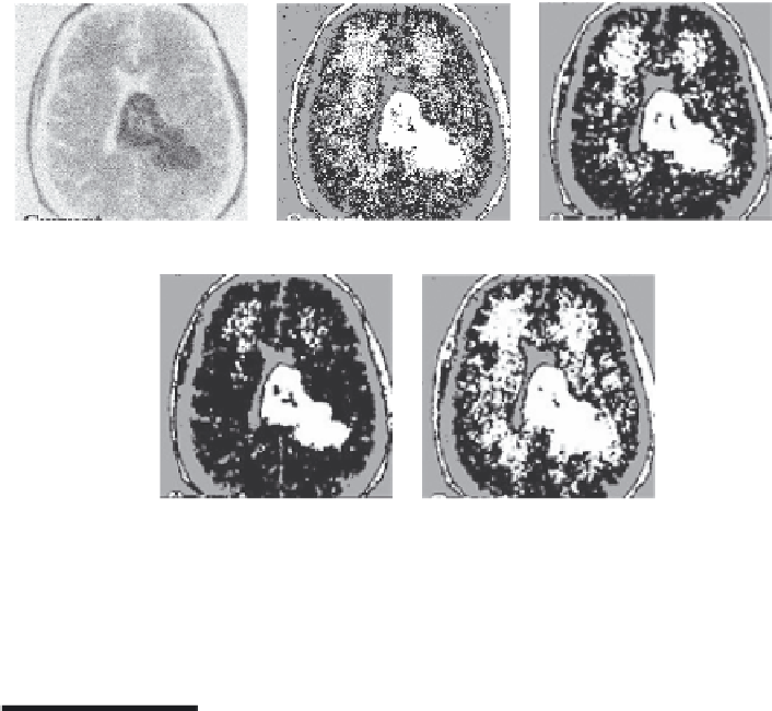
(a)
(b)
(c)
(d)
(e)
FIGURE 7.8
(a) Original image, (b) IFCM, (c) IFCM with Gaussian kernel, (d) IFCM with hypertangent ker-
nel and (e) IFCM with radial basis kernel. (Modified from Chaira, T. and Panwar, A.,
J. Comput.
Intell. Syst.
, 7(2), 1-11, 2013.)
7.8 Colour Clustering
The objective of colour clustering is to divide or cluster the image into sev-
eral homogeneous regions using colour as a feature. Colour approach is an
important issue especially when we are dealing with medical images, for
example, cells or tissues or skin-related problems, where the effect of colour
clustering is analysed to diagnose different types of diseases. Recently, cell
classification has a widespread interest especially in pathological laborato-
ries where blood cell counting of red blood cell (RBC) and white blood cell
(WBC) is carried out for disease detection. Blood cell can be RBC, WBC and
blood platelets, and separating WBC from the blood cell is a very critical task.
The difference in these types lies in the texture, colour of the cytoplasm and
nucleus. In blood smear images, the RBCs are more than the WBCs. Accurate
cell image segmentation for determining valuable quantitative diagnostic
information for pathologists is necessary. Now, with the automated tech-
niques such as medical imaging, analysing becomes faster and gives more
accurate results. But the requirements for clustering pathological images
are different from the general clustering of images as the colours (stained
cell images) are vaguely distributed and so clustering of cells becomes very
Search WWH ::

Custom Search