Biomedical Engineering Reference
In-Depth Information
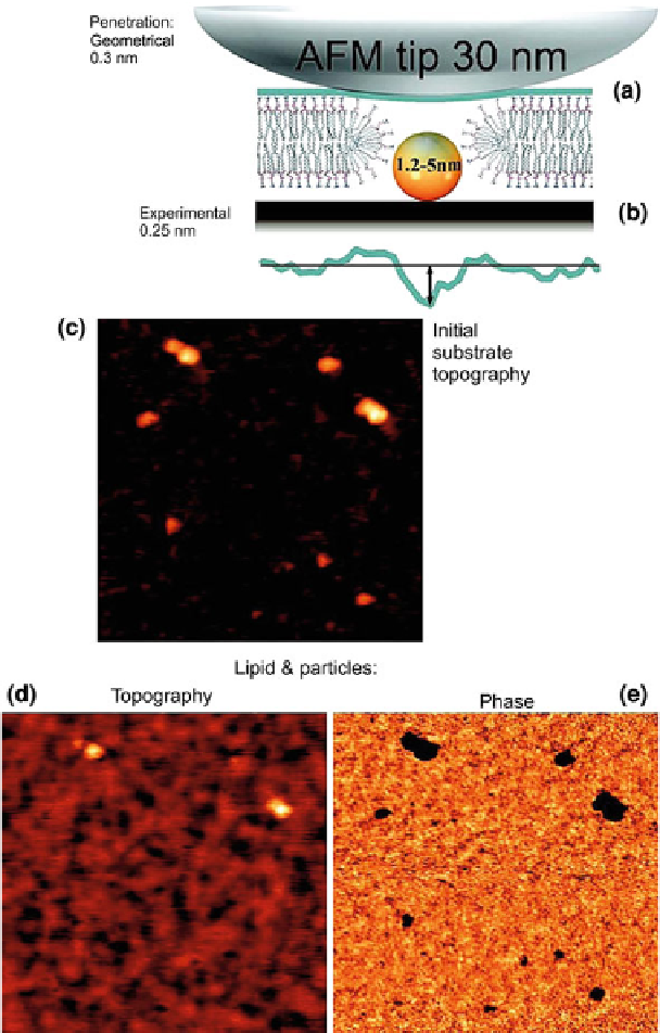
Fig. 6.9
AFM data confirming the formation of pores around 1.2-5nm diameter nanoparticles:
a
theoretical and
b
experimental AFM topography of a pore in the membrane,
c
a topography
image of the substrate with particles (no lipid),
d
topography, and
e
phase imaging of the lipid
deposited on the sample with 1-8nm diameter nanoparticles (scan size 475
475nm
2
). This figure
×
with its description has been taken with the publisher's permission from [
22
]
Search WWH ::

Custom Search